
















©The Author(s) 2025.
World J Hepatol. Dec 27, 2025; 17(12): 113660
Published online Dec 27, 2025. doi: 10.4254/wjh.v17.i12.113660
Published online Dec 27, 2025. doi: 10.4254/wjh.v17.i12.113660
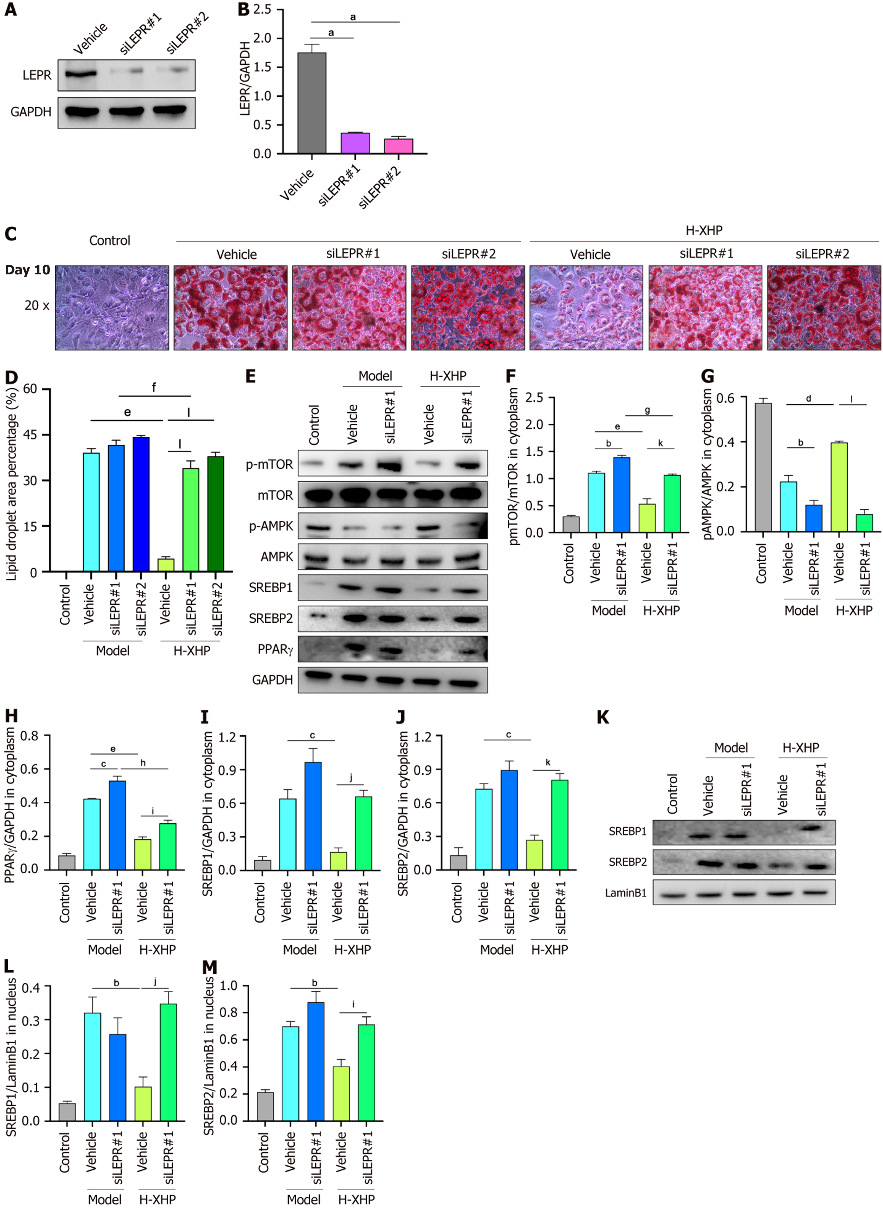
Figure 8 Inhibition of lipid synthesis by Xietu Hemu prescription following siRNA disruption of leptin receptor in 3T3-L1 cells.
A and B: SiRNA disruption of 3T3-L1 cell LEPR efficiency test; C and D: Oil red O staining micro camera and statistics of difference in lipid droplet area occupation on day 10 with/without high dose of Xietu Hemu prescription (H-XHP) or siLEPR intervention; E-J: Detection and quantitative statistics of cytoplasm protein levels on day 10 with/without H-XHP or siLEPR intervention (n = 3); K-M: Detection and quantitative statistics of nucleus protein levels on day 10 with/without H-XHP or siLEPR intervention (n = 3). Data were expressed as mean ± SEM. 200 ×, 50 μm; 400 ×, 25 μm; aP < 0.05 vs vehicle; bP < 0.05 vs model-vehicle; cP < 0.01 vs model-vehicle; dP < 0.005 vs model-vehicle; eP < 0.001 vs model-vehicle; fP < 0.05 vs model-siLEPR1; gP < 0.01 vs model-siLEPR1;hP < 0.001 vs model-siLEPR1; iP < 0.05 vs high dose of Xietu Hemu prescription (H-XHP)-vehicle; jP < 0.01 vs H-XHP-vehicle; kP < 0.005 vs H-XHP-vehicle; lP < 0.001 vs H-XHP-vehicle. H-XHP: High dose of Xietu Hemu prescription.
- Citation: Cheng Z, Lu YF, He YX, Wei W, Xie YX, Lv TS, Wei Y, Lou Y, Yu JY, Zhou XQ. Integrated serum metabolomics reveal molecular mechanism of Xietu Hemu prescription on metabolic dysfunction-associated steatotic liver disease-related obesity. World J Hepatol 2025; 17(12): 113660
- URL: https://www.wjgnet.com/1948-5182/full/v17/i12/113660.htm
- DOI: https://dx.doi.org/10.4254/wjh.v17.i12.113660