
















©The Author(s) 2025.
World J Hepatol. Dec 27, 2025; 17(12): 111414
Published online Dec 27, 2025. doi: 10.4254/wjh.v17.i12.111414
Published online Dec 27, 2025. doi: 10.4254/wjh.v17.i12.111414
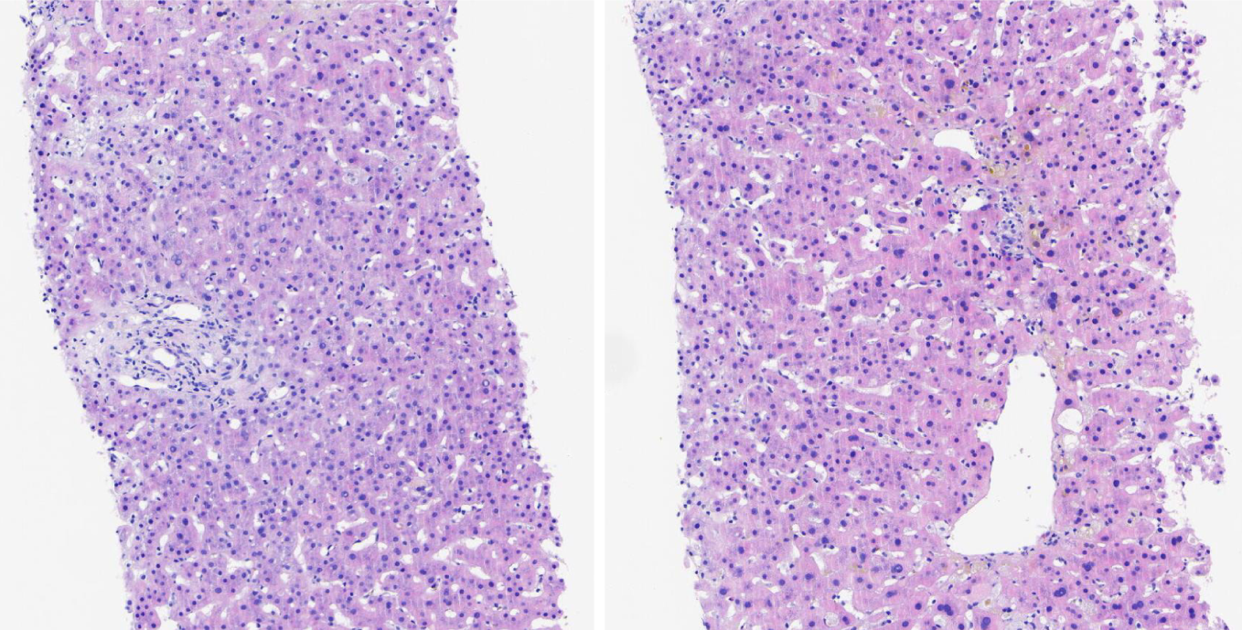
Figure 3 Histological findings of an ultrasound-guided percutaneous liver biopsy stained with hematoxylin and eosin (magnification × 10).
A histological examination revealed severe lobular hepatitis, accompanied by bile duct injury and intrahepatic cholestasis, and a loss of bile duct structure around portal tracts with a small amount of visible lymphocytes and plasma cell infiltration, which was believed to be the result of drug-related liver injury.
- Citation: Xu MJ, Wei X, Lu Y, Sun L, Xie Y, Li MH. Severe hyperlipidemia associated with drug-induced cholestatic liver disease: A case report. World J Hepatol 2025; 17(12): 111414
- URL: https://www.wjgnet.com/1948-5182/full/v17/i12/111414.htm
- DOI: https://dx.doi.org/10.4254/wjh.v17.i12.111414