
















©The Author(s) 2025.
World J Hepatol. Dec 27, 2025; 17(12): 110733
Published online Dec 27, 2025. doi: 10.4254/wjh.v17.i12.110733
Published online Dec 27, 2025. doi: 10.4254/wjh.v17.i12.110733
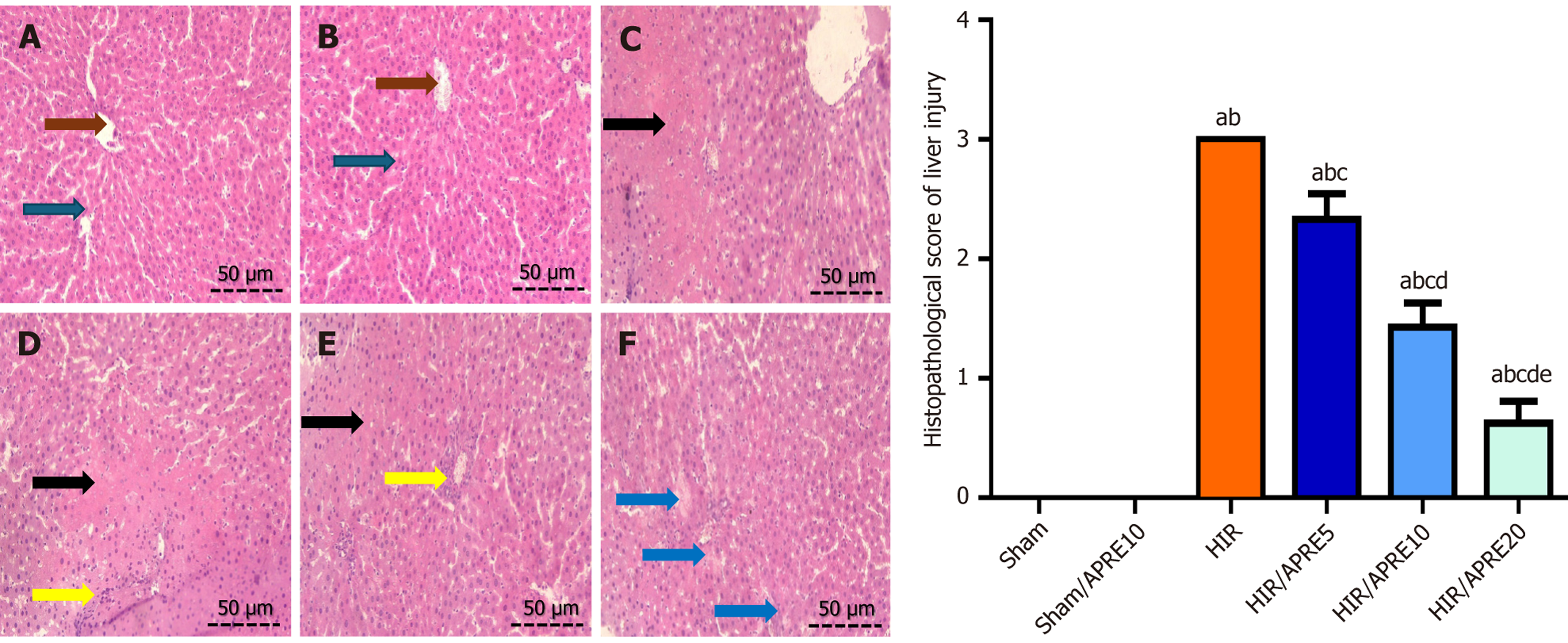
Figure 4 Histopathology of hepatic tissues in hepatic ischemia reperfusion-induced liver injury in rats.
A and B: Slides of both Sham (A) and Sham/aprepitant (APRE) 10 group (B) groups showed a normal central vein (brown arrows) lined by endothelial cells with normal polygonal hepatocytes (blue arrows); C: Slides of the hepatic ischemia reperfusion (HIR) group showed marked centrilobular necrosis (black arrow); D: Showed moderate hepatocyte necrosis (black arrow), degeneration, and inflammatory cell infiltration (yellow arrow). E: Slides of the HIR/APRE10 group showed mild hepatocyte necrosis (black arrow), degeneration, and inflammatory cell infiltrate (yellow arrow); F: The HIR/APRE20 group. Results are considered significantly different when P < 0.05. aP < 0.05 significantly different from Sham group; bP < 0.05 significantly different from Sham/aprepitant (APRE) 10; cP < 0.05 significantly different from hepatic ischemia reperfusion (HIR) group; dP < 0.05 significantly different from HIR/APRE5; eP < 0.05 significantly different from HIR/APRE10 group. APRE5: Aprepitant 5 mg/kg; APRE10: Aprepitant 10 mg/kg; APRE20: Aprepitant 20 mg/kg; HIR: Hepatic ischemia reperfusion.
- Citation: Kelleni MT, Abdelzaher WY, Adly M, Attya ME, Fawzy MA, Ibrahim MA. Targeting sirtuin 1/nuclear factor erythroid 2-related factor 2/tumor necrosis factor-α pathway to modulate hepatic ischemia reperfusion-induced injury. World J Hepatol 2025; 17(12): 110733
- URL: https://www.wjgnet.com/1948-5182/full/v17/i12/110733.htm
- DOI: https://dx.doi.org/10.4254/wjh.v17.i12.110733