
















©The Author(s) 2025.
World J Hepatol. Nov 27, 2025; 17(11): 110050
Published online Nov 27, 2025. doi: 10.4254/wjh.v17.i11.110050
Published online Nov 27, 2025. doi: 10.4254/wjh.v17.i11.110050
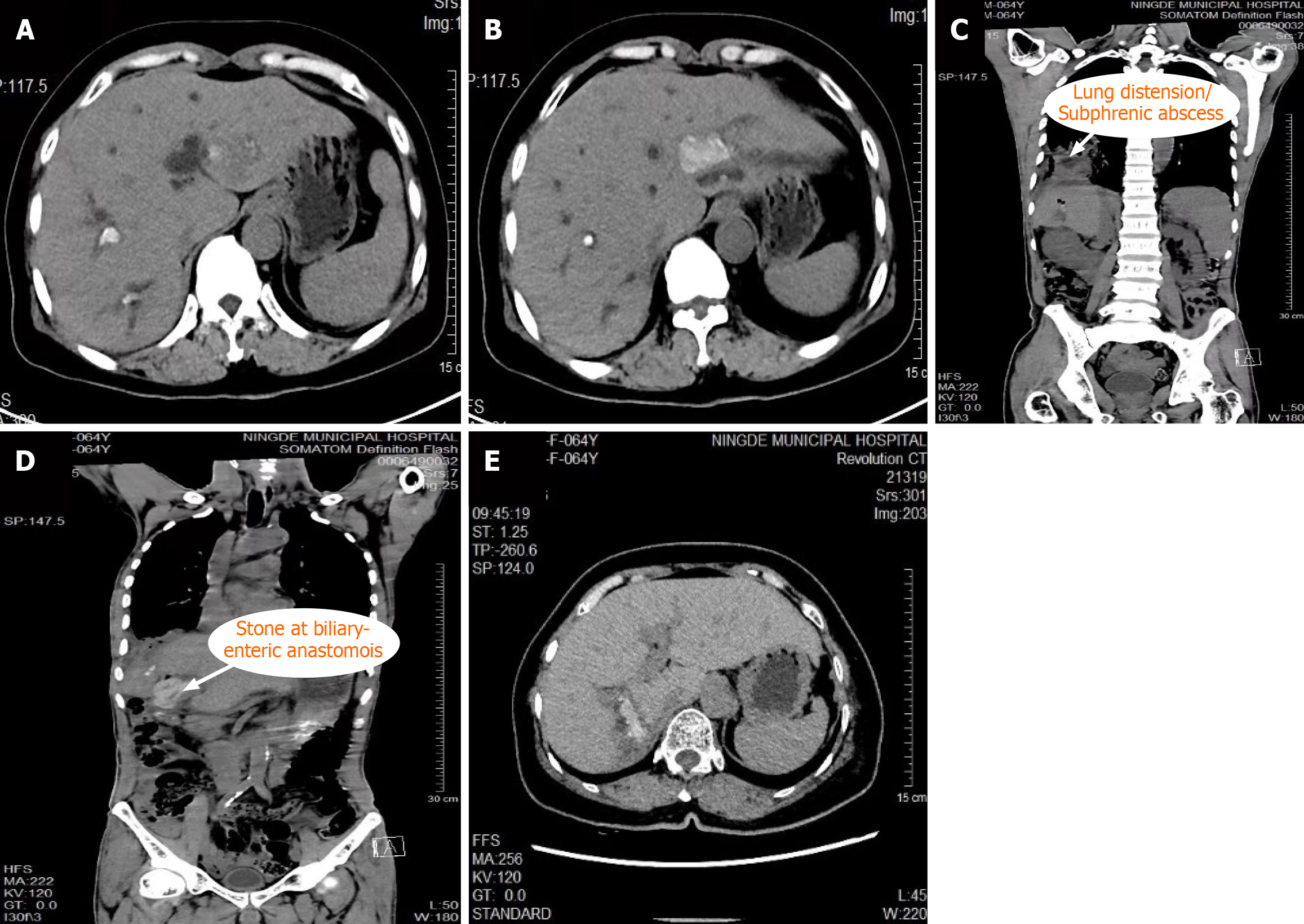
Figure 1 Preoperative images of selected cases with complex hepatolithiasis.
A and B: Preoperative axial and coronal computed tomography (CT) scans showing diffuse intrahepatic biliary stones in the left and right hepatic lobes; C: CT scan demonstrating lung expansion with subphrenic abscess secondary to recurrent cholangitis; D: CT scan showing a retained stone at the site of prior biliary-enteric anastomosis; E: CT scan revealing severe intrahepatic bile duct dilatation accompanied by parenchymal atrophy.
- Citation: Lin DX, Zhuo XB, Chang GJ, Lei WD, Huang J, Zhang Y, Qiu ZJ, Zhang SY. Laparoscopic vs open surgery for complex hepatolithiasis: A retrospective comparative study. World J Hepatol 2025; 17(11): 110050
- URL: https://www.wjgnet.com/1948-5182/full/v17/i11/110050.htm
- DOI: https://dx.doi.org/10.4254/wjh.v17.i11.110050