
















©The Author(s) 2025.
World J Hepatol. Oct 27, 2025; 17(10): 110849
Published online Oct 27, 2025. doi: 10.4254/wjh.v17.i10.110849
Published online Oct 27, 2025. doi: 10.4254/wjh.v17.i10.110849
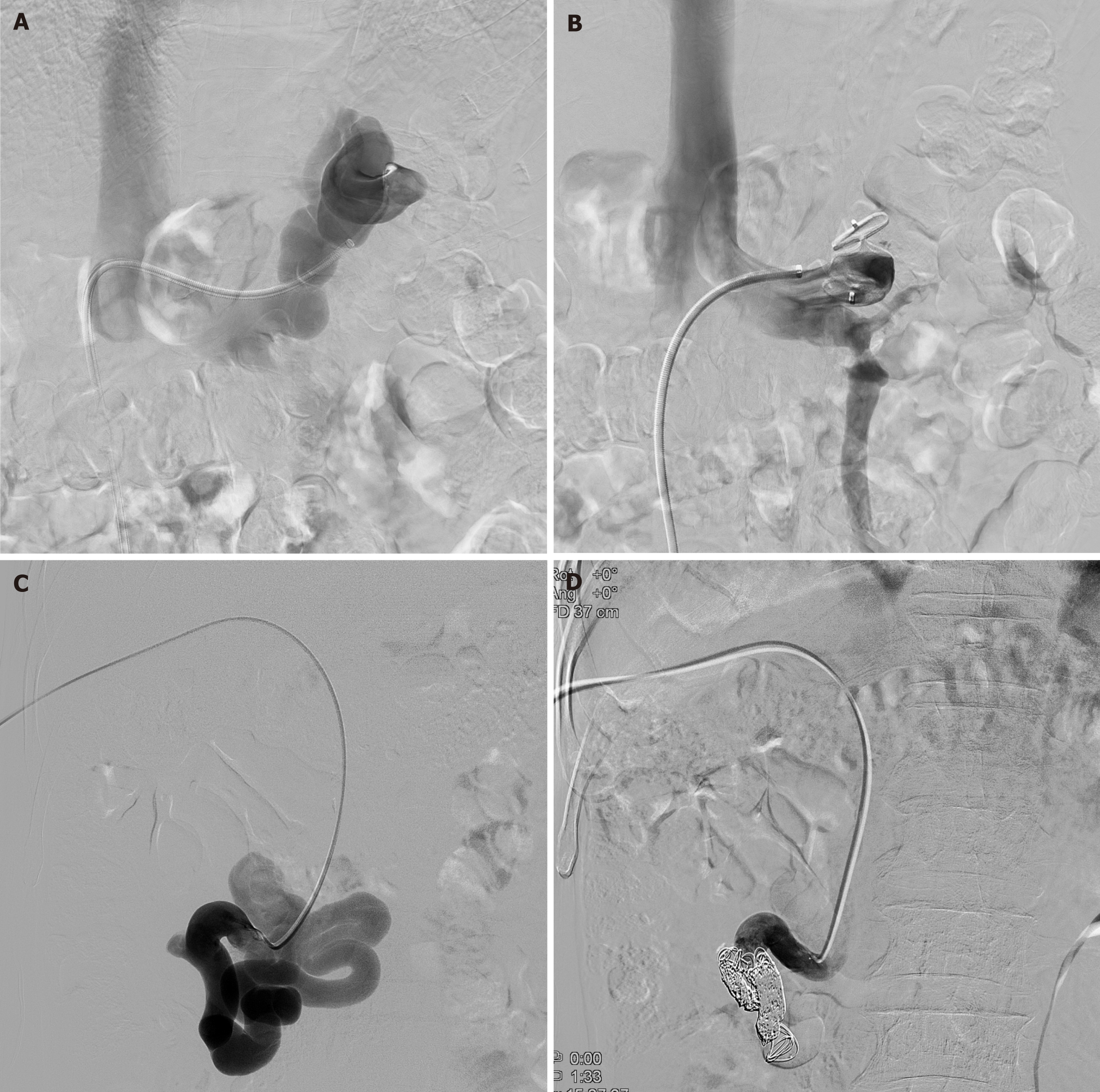
Figure 3 Angiographic images of splenorenal shunt embolization and mesocaval shunt embolization.
A: After a 9-F vascular sheath was placed in the splenorenal shunt, the shunt was accessed coaxially using a 5-F cobra catheter; B: The vascular plug was deployed at the narrowest region of the splenorenal shunt through the vascular sheath and completion venography showed complete occlusion of the splenorenal shunt; C: Transhepatic superior mesenteric venogram showed the large size of mesocaval shunt; D: Coil placement showed complete closure of mesocaval shunt.
- Citation: Park JW, Kim Y, Lee JS, Jung IS, Kim KB, Chae HB. Improved clinical outcomes following embolization of extrahepatic portosystemic shunts in cirrhotic patients with recurrent hepatic encephalopathy. World J Hepatol 2025; 17(10): 110849
- URL: https://www.wjgnet.com/1948-5182/full/v17/i10/110849.htm
- DOI: https://dx.doi.org/10.4254/wjh.v17.i10.110849