
















©The Author(s) 2025.
World J Hepatol. Oct 27, 2025; 17(10): 110029
Published online Oct 27, 2025. doi: 10.4254/wjh.v17.i10.110029
Published online Oct 27, 2025. doi: 10.4254/wjh.v17.i10.110029
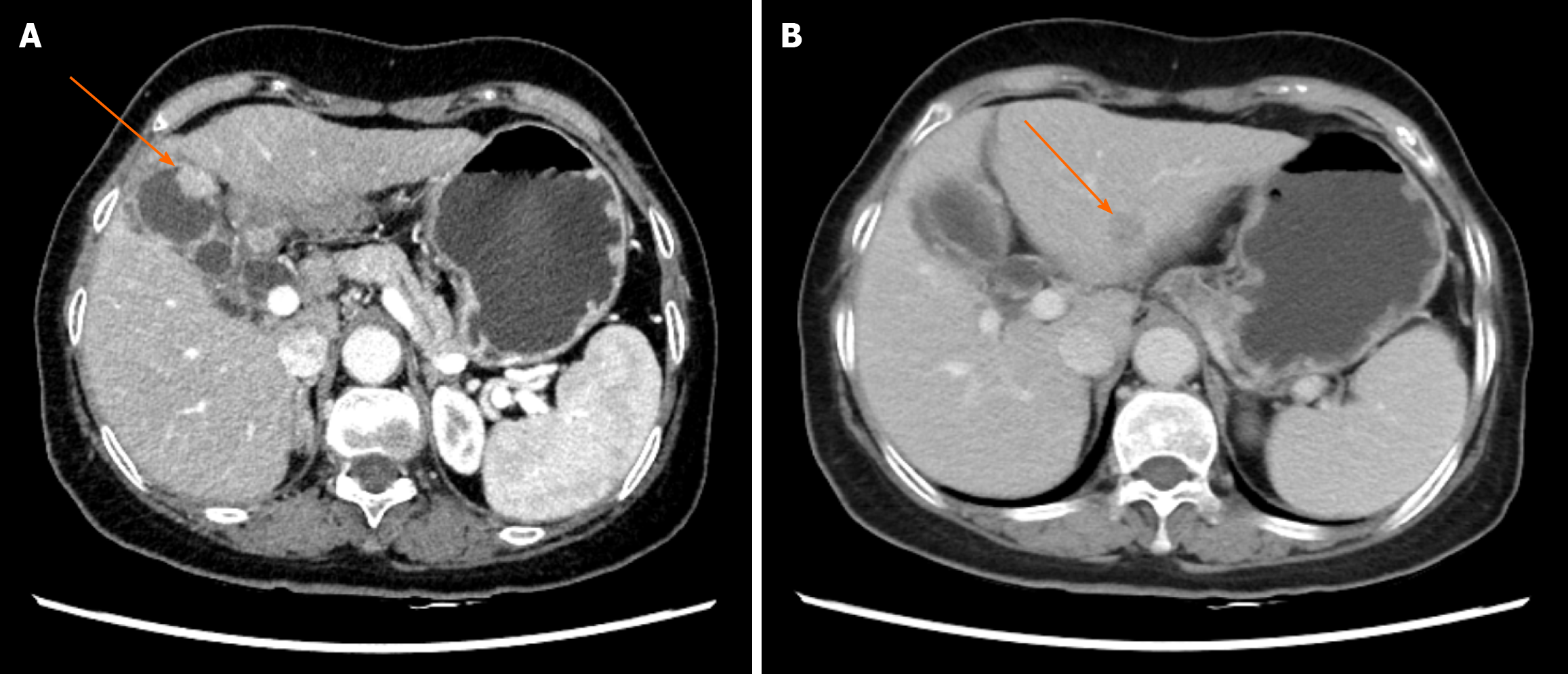
Figure 1 Abdominal computed tomography.
A: Abdominal computed tomography showing a 2.0 cm × 1.0 cm mass in the gallbladder. Arrow: Gallbladder mass (axial view); B: Abdominal computed tomography showing a 1.6 cm × 1.5 cm lesion in the left lateral lobe of the liver. Arrow: Liver lesion (axial view).
- Citation: Zhang K, Liu HL. Unusual presentation of synchronous double primary gallbladder and hepatic malignancies: A case report. World J Hepatol 2025; 17(10): 110029
- URL: https://www.wjgnet.com/1948-5182/full/v17/i10/110029.htm
- DOI: https://dx.doi.org/10.4254/wjh.v17.i10.110029