
















©The Author(s) 2025.
World J Hepatol. Oct 27, 2025; 17(10): 110026
Published online Oct 27, 2025. doi: 10.4254/wjh.v17.i10.110026
Published online Oct 27, 2025. doi: 10.4254/wjh.v17.i10.110026
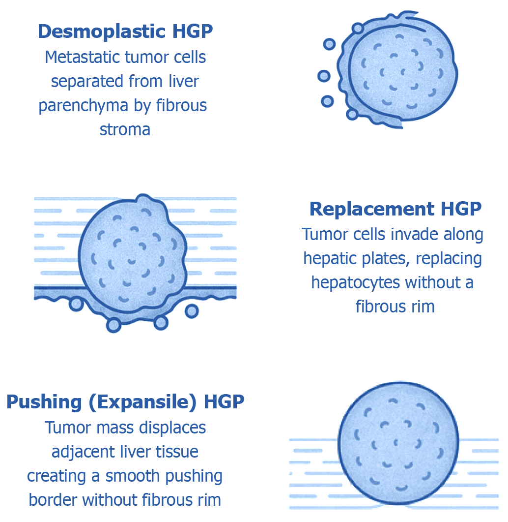
Figure 1 Histological growth patterns at the tumor–liver interface in hepatic metastases.
This diagram depicts the three major histopathological growth patterns (HGPs) that characterize how metastatic tumors interact with the surrounding liver parenchyma, each representing a distinct tumor–microenvironment relationship: (1) Desmoplastic pattern: Defined by the presence of a dense fibrotic stromal band encircling the tumor, effectively separating malignant cells from the adjacent hepatic tissue. This interface is commonly enriched with immune cell infiltrates and newly formed blood vessels, reflecting active stromal remodeling; (2) Replacement pattern: In this growth mode, tumor cells infiltrate the liver parenchyma by replacing hepatocytes along existing sinusoidal structures. The architecture of the liver is largely preserved, and no intervening fibrotic stroma is present. Tumor cells often hijack the native hepatic vasculature to support their growth; and (3) Pushing (expansile) pattern: Characterized by a sharply defined tumor boundary that displaces, rather than infiltrates, the surrounding liver tissue. This interface is smooth and lacks both fibrous stroma and invasion into adjacent hepatocytes. These histological patterns are not only morphologically distinct but also carry significant prognostic and biological implications, influencing metastatic progression, response to therapy, and overall clinical outcomes. HGP: Histopathological growth patterns.
- Citation: Ebrahim NAA, Farghaly TA, El-Sherif AA, Fahmy AM, Othman MO, Tahoun NS, Korany OM, Arafat A, Oreaba R, Soliman SMA. Clinicopathological insights and management of liver metastases: Current advances and future perspectives. World J Hepatol 2025; 17(10): 110026
- URL: https://www.wjgnet.com/1948-5182/full/v17/i10/110026.htm
- DOI: https://dx.doi.org/10.4254/wjh.v17.i10.110026