
















©The Author(s) 2025.
World J Hepatol. Oct 27, 2025; 17(10): 109575
Published online Oct 27, 2025. doi: 10.4254/wjh.v17.i10.109575
Published online Oct 27, 2025. doi: 10.4254/wjh.v17.i10.109575
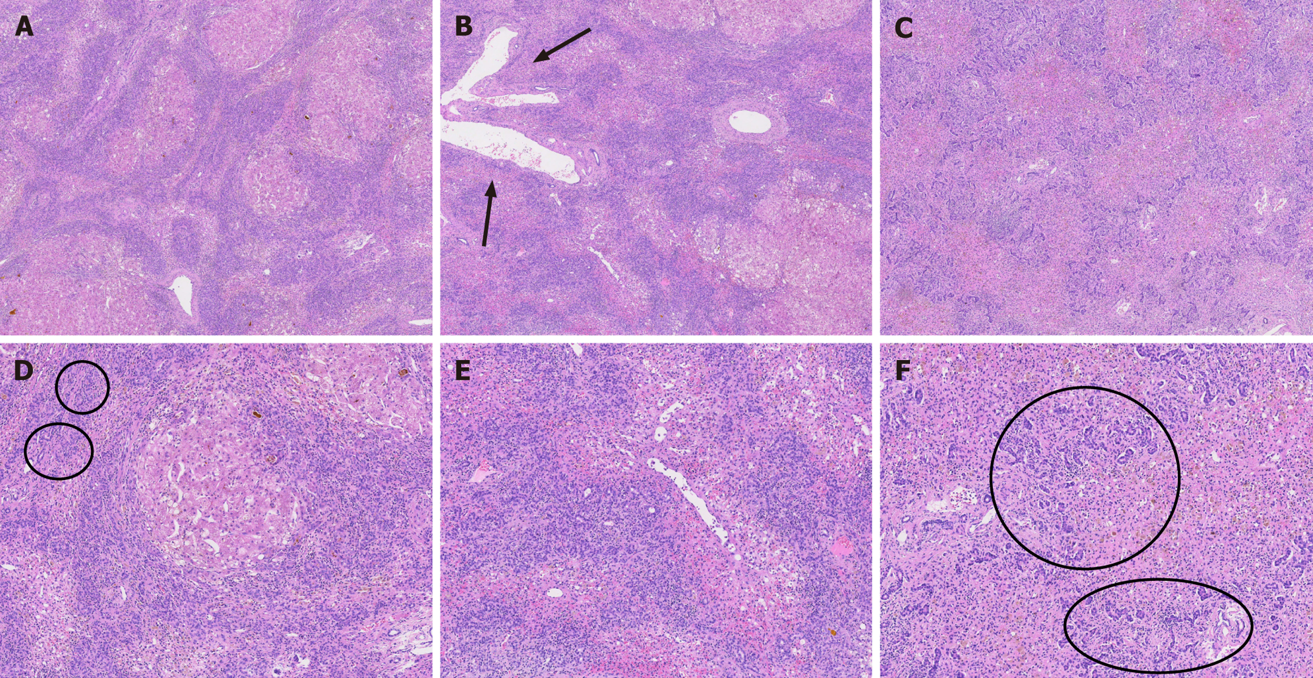
Figure 3 Postoperative pathology.
A: Severe interface hepatitis: Massive lymphocytic infiltration, hematoxylin and eosin (HE) stained, 4 ×; B: Portal inflammation: Lymphocytic infiltration in the portal area, with markedly dilated portal vein branches (arrow indicates dilated portal branches), HE, 4 ×; C: Inflammatory infiltrate: Predominantly lymphocytes, HE, 4 ×; D: Bile thrombus formation (circled area shows bile thrombi), HE, 10 ×; E: Sinusoidal dilatation (Disse space), HE, 10 ×; F: Dense bile ductular proliferation (circled area shows clustered proliferating bile ductules), HE, 10 ×.
- Citation: Zhu XY, Zhao YT, Su CS, Yuan XD, Zhang SG, Nashan B. Acute liver failure caused by alkaloids from traditional Chinese medicine: A case report. World J Hepatol 2025; 17(10): 109575
- URL: https://www.wjgnet.com/1948-5182/full/v17/i10/109575.htm
- DOI: https://dx.doi.org/10.4254/wjh.v17.i10.109575