
















©The Author(s) 2025.
World J Hepatol. Oct 27, 2025; 17(10): 109517
Published online Oct 27, 2025. doi: 10.4254/wjh.v17.i10.109517
Published online Oct 27, 2025. doi: 10.4254/wjh.v17.i10.109517
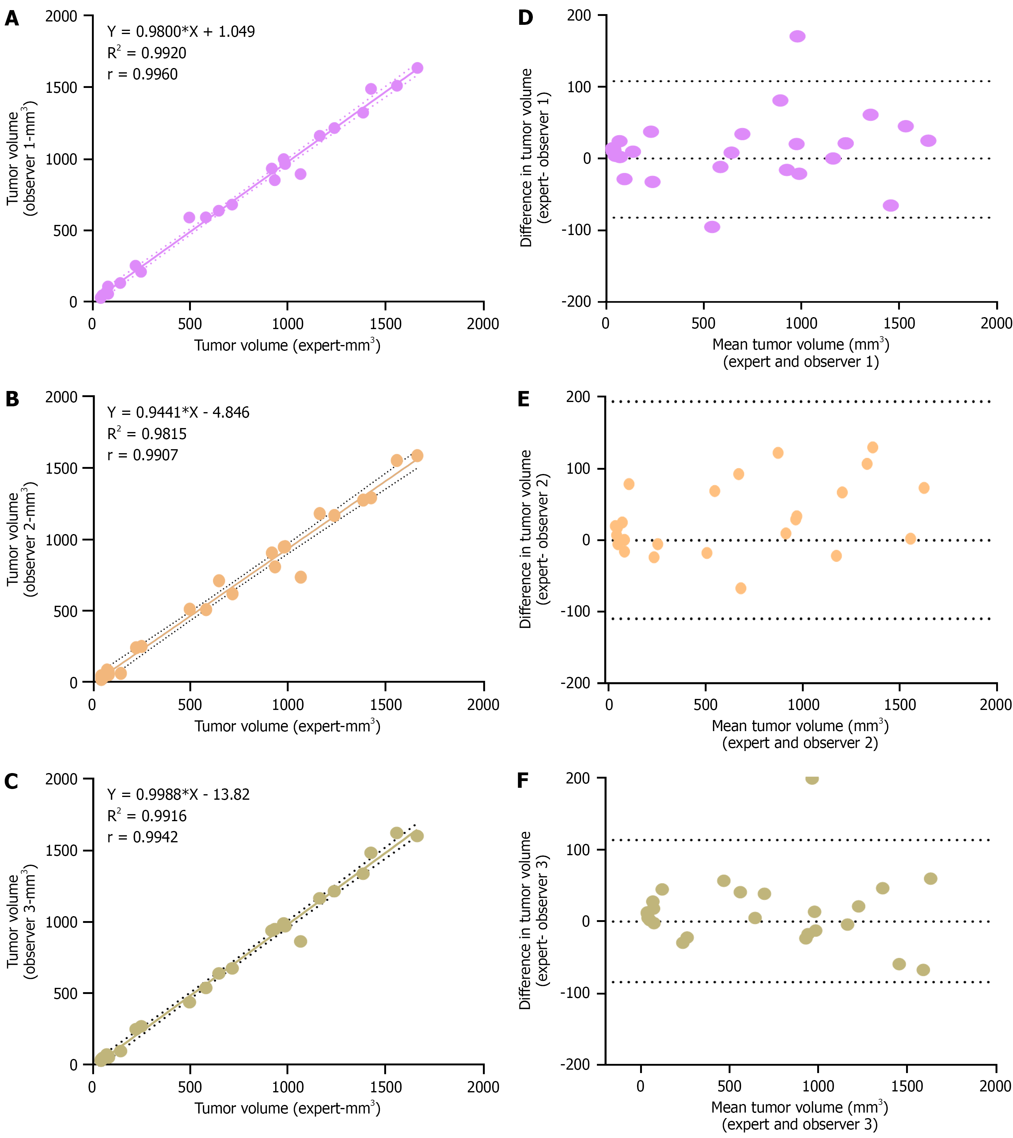
Figure 4 Evaluation of expert non-expert variability in tumor volume measurement by ultrasound imaging.
A-C: A linear correlation plot showing the association of tumour volume measurement by observer 1, 2 and 3 respectively with expert measurement. Dotted line indicates 95% confidence bands and Pearson correlation coefficient is mentioned in each plot (P < 0.001, n = 25 for each plot); D-F: Bland-Altman plots showing the deviation in observer measurements from expert measurements. Upper and lower dotted lines represent the limits of agreement.
- Citation: Devan AR, Sasidharan SM, Sreekumar KP, Unni AKK, Mangalathillam S, Ansar A, Unni AR, Nath LR. Ultrasound imaging-guided protocol for monitoring tumor growth in orthotopic rat model of hepatocellular carcinoma. World J Hepatol 2025; 17(10): 109517
- URL: https://www.wjgnet.com/1948-5182/full/v17/i10/109517.htm
- DOI: https://dx.doi.org/10.4254/wjh.v17.i10.109517