
















©The Author(s) 2025.
World J Hepatol. Oct 27, 2025; 17(10): 109517
Published online Oct 27, 2025. doi: 10.4254/wjh.v17.i10.109517
Published online Oct 27, 2025. doi: 10.4254/wjh.v17.i10.109517
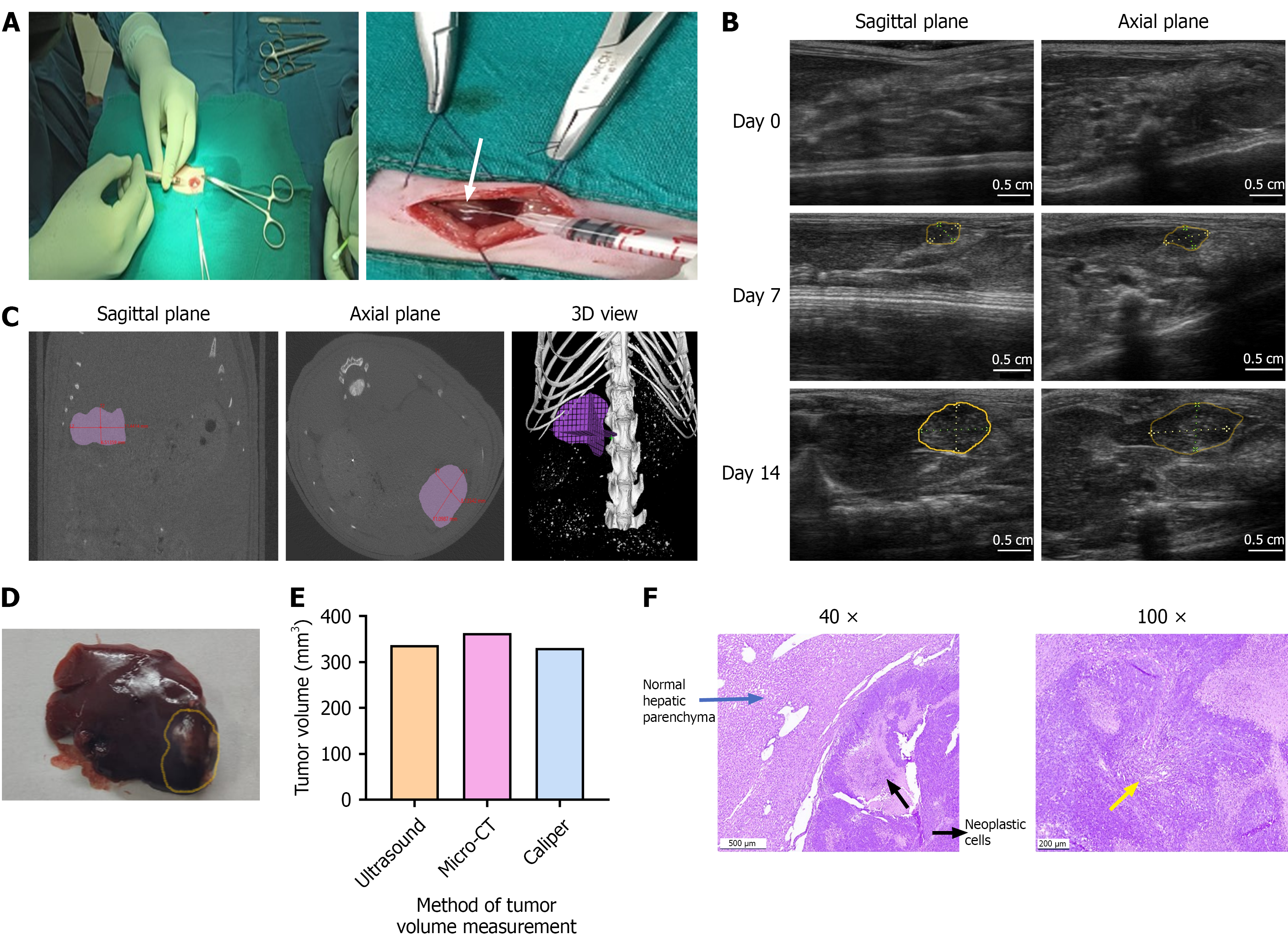
Figure 1 Surgical induction of hepatocellular carcinoma and validation of ultrasound imaging for tumor monitoring in comparison with micro-computed tomography.
A: Hepatocellular carcinoma (HCC) was induced by injecting 5 × 10⁶ N1S1 cells into the subcapsular region at the tip of the left lateral liver lobe of Sprague-Dawley rats. The formation of a white bleb on the liver surface (white arrow) confirmed successful cell implantation. The tumor induction rate was 100%, with a single, well-defined hypoechoic nodule developing in all animals by day 7 post-implantation; B: Ultrasound (US) imaging of the liver was performed in both sagittal and axial planes prior to cell injection (day 0) and weekly thereafter (days 7 and 14). Tumor formation was confirmed by day 7, with evident tumor progression by day 14 (tumor area outlined in yellow); C: Micro-computed tomography (micro-CT) imaging was performed on day 14 and analysed in planes (sagittal and axial) similar to those used for US imaging. Tumor dimensions were measured and validated with 3D view. micro-CT confirmed the presence of a growing tumor with volume measurements comparable to those obtained by US; D: Gross liver morphology on day 14 revealed a tumor nodule at the tip of the left lateral lobe (tumor area outlined in yellow), with tumor volume measured using a vernier caliper (VC); E: Comparative tumor volume measurements from US, CT and VC in a representative rat bearing orthotopic N1S1 HCC tumor. All imaging and measurements were performed on day 14 post-implantation; F: Representative histology images of H&E staining in the liver tumor tissues of SD rats. Images include SD rat HCC tumor tissue on day 14 at 40 × and 100 × magnification (size bar for: For: 500 μm = 40 ×, 200 μm = 100 ×). Histopathological analysis confirmed mid-stage HCC consists of dense proliferating neoplastic cells (yellow arrow) invading into the adjacent normal hepatic parenchyma.
- Citation: Devan AR, Sasidharan SM, Sreekumar KP, Unni AKK, Mangalathillam S, Ansar A, Unni AR, Nath LR. Ultrasound imaging-guided protocol for monitoring tumor growth in orthotopic rat model of hepatocellular carcinoma. World J Hepatol 2025; 17(10): 109517
- URL: https://www.wjgnet.com/1948-5182/full/v17/i10/109517.htm
- DOI: https://dx.doi.org/10.4254/wjh.v17.i10.109517