
















©The Author(s) 2024.
World J Hepatol. Feb 27, 2024; 16(2): 279-285
Published online Feb 27, 2024. doi: 10.4254/wjh.v16.i2.279
Published online Feb 27, 2024. doi: 10.4254/wjh.v16.i2.279
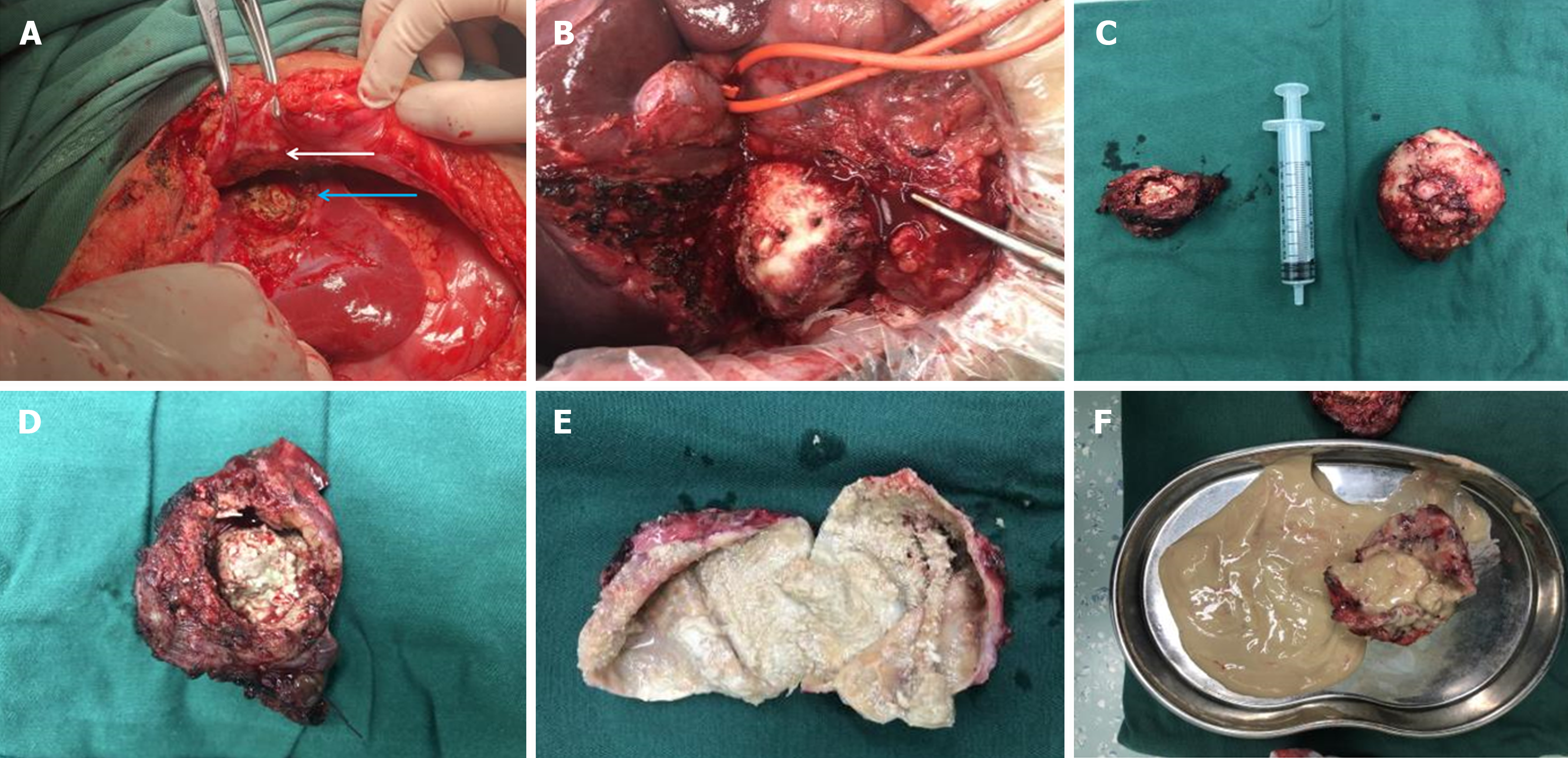
Figure 3 Intraoperative pathology specimens.
A and B: Intraoperative visible lesions; C: Intraoperative excision of pathologic specimens (hepatic cystic echinococcosis and hepatic alveolar echinococcosis); D: Intraoperative excision of pathologic specimens (hepatic alveolar echinococcosis); E: Intraoperative excision of pathologic specimens (hepatic cystic echinococcosis); F: Intraoperative excision of pathologic specimens. Note: The white arrows indicate the site of the abdominal wall abscess sinus tract, and the blue arrows indicate the site of the hepatic alveolar echinococcosis lesion.
- Citation: Wang MM, An XQ, Chai JP, Yang JY, A JD, A XR. Coinfection with hepatic cystic and alveolar echinococcosis with abdominal wall abscess and sinus tract formation: A case report. World J Hepatol 2024; 16(2): 279-285
- URL: https://www.wjgnet.com/1948-5182/full/v16/i2/279.htm
- DOI: https://dx.doi.org/10.4254/wjh.v16.i2.279