
















©The Author(s) 2021.
World J Hepatol. Sep 27, 2021; 13(9): 1079-1097
Published online Sep 27, 2021. doi: 10.4254/wjh.v13.i9.1079
Published online Sep 27, 2021. doi: 10.4254/wjh.v13.i9.1079
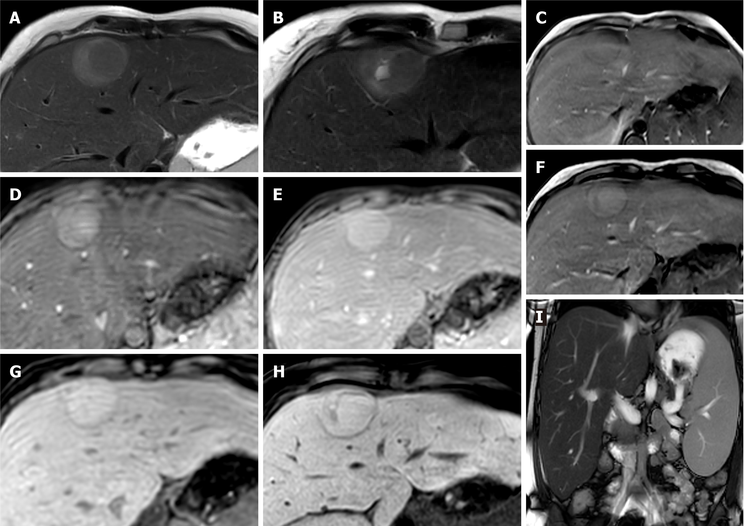
Figure 2 Hepatocellular adenoma.
A 27-year-old lady with background of glycogen storage type 1 disease. A and B: Segment IVA liver lesion demonstrating mild T2 hyperintensity with atoll sign (A) and cystic foci (B); C and F: No signal drop out on out-of-phase (F) when compared to in-phase (C) T1-weighted sequence; D, E and G: There is quite homogenous hyperenhancement on arterial phase (D) with no washout on portal venous (E) and delayed (G) phases; H: Hepatobiliary phase shows contrast retention within the lesion; I: Coronal T2-weighted shows hepatosplenomegaly as features of glycogen storage disease type I. The lesion has increased in size and therefore was resected, histology revealed an inflammatory subtype hepatocellular adenoma.
- Citation: Noreikaite J, Albasha D, Chidambaram V, Arora A, Katti A. Indeterminate liver lesions on gadoxetic acid-enhanced magnetic resonance imaging of the liver: Case-based radiologic-pathologic review. World J Hepatol 2021; 13(9): 1079-1097
- URL: https://www.wjgnet.com/1948-5182/full/v13/i9/1079.htm
- DOI: https://dx.doi.org/10.4254/wjh.v13.i9.1079