Copyright
©The Author(s) 2021.
World J Hepatol. May 27, 2021; 13(5): 584-594
Published online May 27, 2021. doi: 10.4254/wjh.v13.i5.584
Published online May 27, 2021. doi: 10.4254/wjh.v13.i5.584
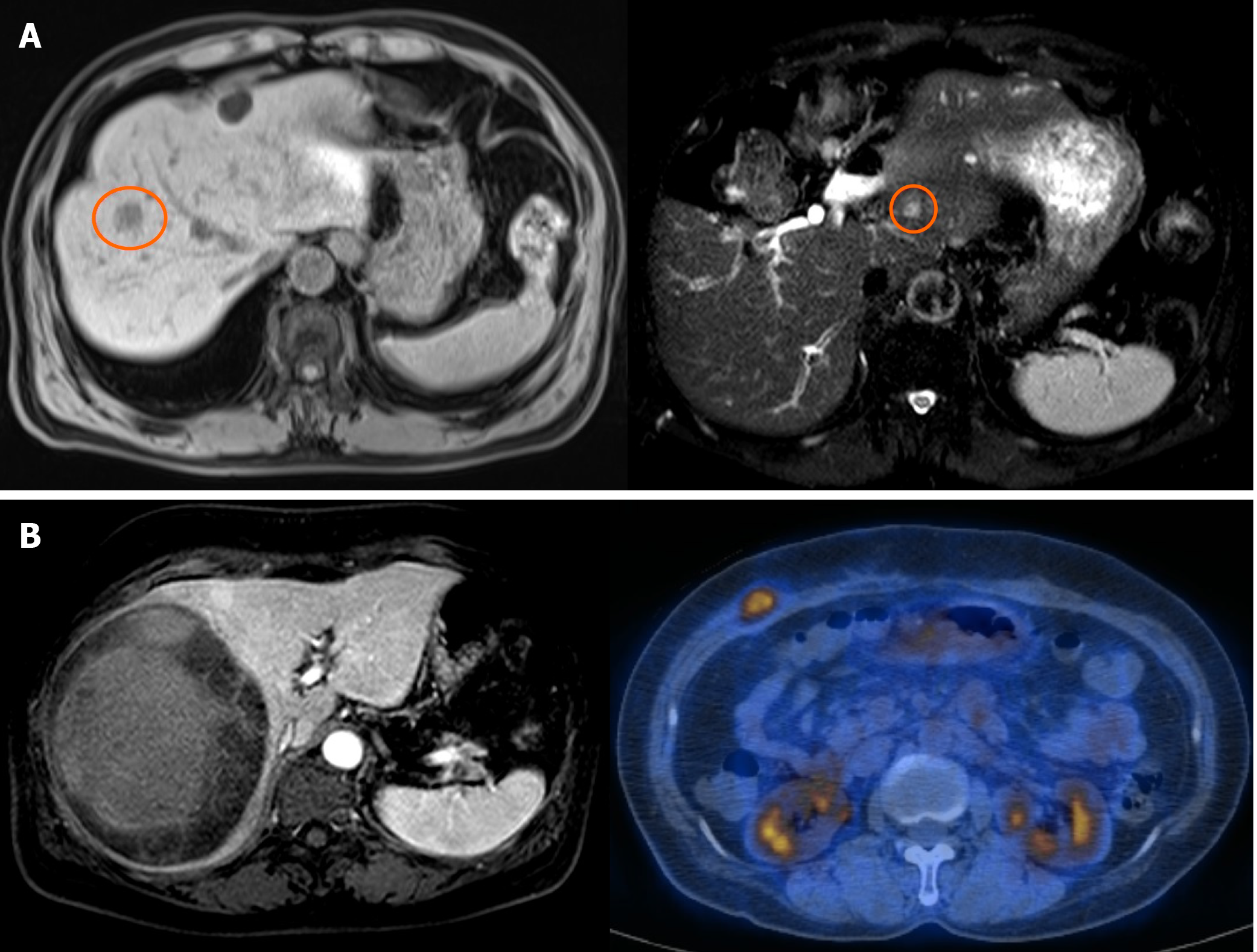
Figure 2 Images of a surviving patient (cases 7 and 8).
A: Case 7. Left: T1 magnetic resonance imaging (MRI) of pre-operative angiosarcoma on S8 (orange circle). Right: T2 MRI of recurrence on S2 five years after central hepatectomy; B: Case 8. Left: MRI of pre-operative sarcoma. An 11.5 cm circumscribed mass with hemorrhage on the Rt. lobe. Right: Positron emission tomography-computed tomography of the recurrent mass. Focal fluoro-deoxyglucose uptake at the Rt. anterior abdominal wall.
- Citation: Kim SJ, Rhu J, Kim JM, Choi GS, Joh JW. Surgical treatment outcomes of primary hepatic sarcomas: A single-center experience. World J Hepatol 2021; 13(5): 584-594
- URL: https://www.wjgnet.com/1948-5182/full/v13/i5/584.htm
- DOI: https://dx.doi.org/10.4254/wjh.v13.i5.584


