Copyright
©The Author(s) 2021.
World J Hepatol. Mar 27, 2021; 13(3): 328-342
Published online Mar 27, 2021. doi: 10.4254/wjh.v13.i3.328
Published online Mar 27, 2021. doi: 10.4254/wjh.v13.i3.328
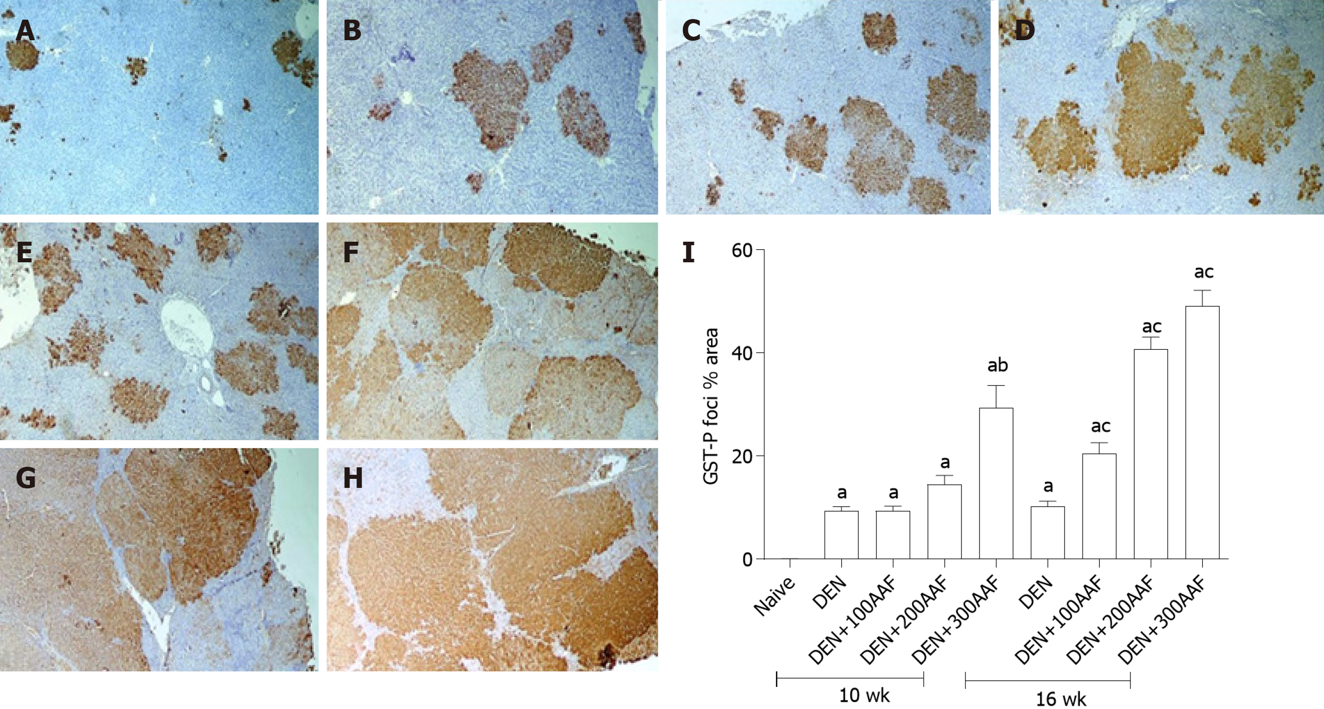
Figure 3 Histological and immunohistochemical examination.
A-H: Images of rats' liver sections immunohistochemically-stained with glutathione S transferase-P (GST-P) antibody, show multiple GST-P-positive hepatic foci and nodules (brown stained collection of cells) of different sizes scatter in-between negatively stained hepatic parenchyma. Rats sacrificed at week 10 (A-D), rats sacrificed at week 16 (E-H) [A and E: diethylnitrosamine (DEN) group; B and F: DEN + 100mg 2-acetylaminofluorene (2-AAF) group; C and G: DEN + 200 mg 2-AAF group; D and H: DEN + 300 mg 2-AAF (× 40)]; I: shows the effect of DEN and 2-AAF at different doses on GSTP foci % area in the liver. Values are mean ± SE; number of animals = 6 rats/each group. aP < 0.05 compared to naïve group; bP < 0.05 compared to DEN group at week 10, cP < 0.05 compared to DEN at week 16 group. One-way ANOVA followed by Tukey's multiple comparison test. DEN: Diethylnitrosamine; 2-AAF: 2-Acetylaminofluorene; GST-P: Glutathione S transferase-P.
- Citation: Hasanin AH, Habib EK, El Gayar N, Matboli M. Promotive action of 2-acetylaminofluorene on hepatic precancerous lesions initiated by diethylnitrosamine in rats: Molecular study. World J Hepatol 2021; 13(3): 328-342
- URL: https://www.wjgnet.com/1948-5182/full/v13/i3/328.htm
- DOI: https://dx.doi.org/10.4254/wjh.v13.i3.328


