Copyright
©The Author(s) 2021.
World J Hepatol. Dec 27, 2021; 13(12): 2192-2200
Published online Dec 27, 2021. doi: 10.4254/wjh.v13.i12.2192
Published online Dec 27, 2021. doi: 10.4254/wjh.v13.i12.2192
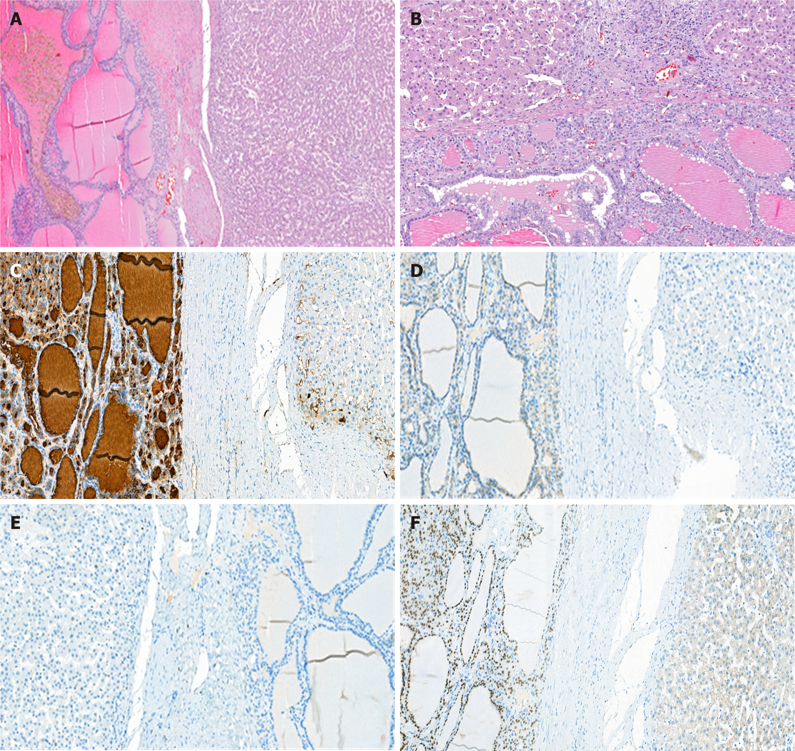
Figure 3 Pathology findings of liver mass.
A: Microscopic appearance - the liver node, with shaped borders, is formed from cavities of different sizes filled with eosinophilic fluid, resembling a colloid (100×); B: Cubic single-layered epithelium lining the cavities (200×). Along the apical surface of the cells, there are characteristic vacuoles in the thick colloid; C: Epithelium labeled with anti-thyroglobulin (2H11 + 6 E1) revealing the thyroid origin (200×); D: Membrane CD56 reveals the neuroendocrine nature of tumor cells (200×); E: A single cell within a tumor node labeled with Ki67, the same as the adjacent normal liver (200×); F: Nuclear TTF-1 immunostaining also suggests a thyroid and thyroid-derived tumor origin (200×).
- Citation: Kovalenko YA, Zharikov YO, Kiseleva YV, Goncharov AB, Shevchenko TV, Gurmikov BN, Kalinin DV, Zhao AV. Rare primary mature teratoma of the liver: A case report. World J Hepatol 2021; 13(12): 2192-2200
- URL: https://www.wjgnet.com/1948-5182/full/v13/i12/2192.htm
- DOI: https://dx.doi.org/10.4254/wjh.v13.i12.2192


