Copyright
©The Author(s) 2021.
World J Hepatol. Dec 27, 2021; 13(12): 1892-1908
Published online Dec 27, 2021. doi: 10.4254/wjh.v13.i12.1892
Published online Dec 27, 2021. doi: 10.4254/wjh.v13.i12.1892
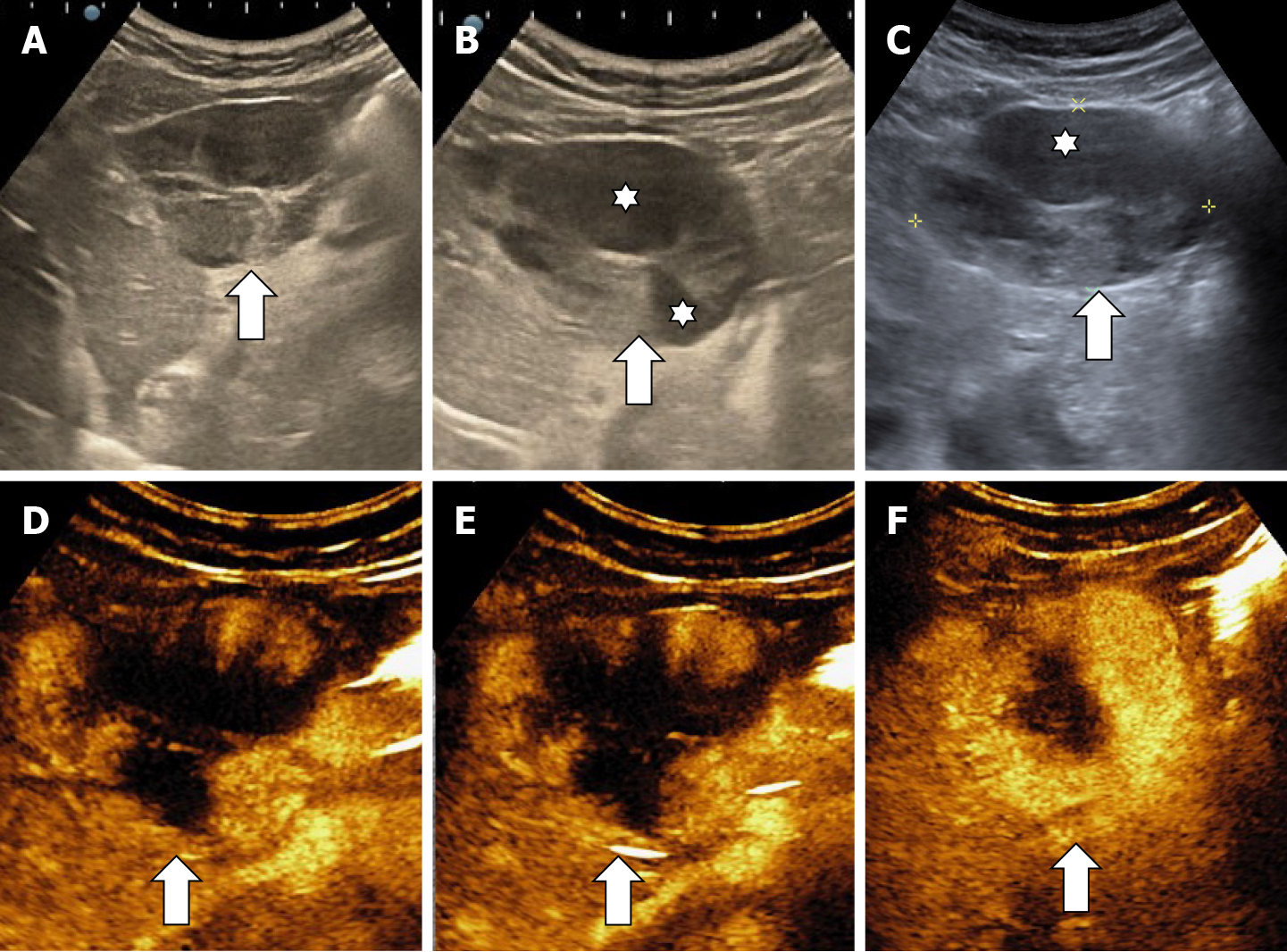
Figure 17 Multicystic hemangioma.
A-C: B mode ultrasound shows an inhomogeneous lesion (A) with central cavity (stars) (B) that contains fluid and septa (C); D-F: In contrast enhanced ultrasound the mass shows a progressive (D and E) but partial filling (F) because of the presence of fluid-like cystic cavities that do not enhance.
- Citation: Sandulescu LD, Urhut CM, Sandulescu SM, Ciurea AM, Cazacu SM, Iordache S. One stop shop approach for the diagnosis of liver hemangioma. World J Hepatol 2021; 13(12): 1892-1908
- URL: https://www.wjgnet.com/1948-5182/full/v13/i12/1892.htm
- DOI: https://dx.doi.org/10.4254/wjh.v13.i12.1892


