Copyright
©The Author(s) 2021.
World J Hepatol. Oct 27, 2021; 13(10): 1450-1458
Published online Oct 27, 2021. doi: 10.4254/wjh.v13.i10.1450
Published online Oct 27, 2021. doi: 10.4254/wjh.v13.i10.1450
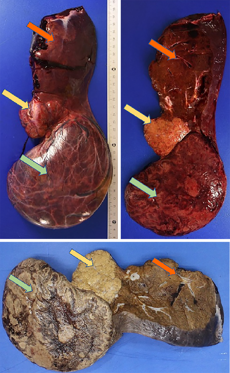
Figure 6 Anatomopathological pictures (top: fresh sample; bottom: formalin-fixed sample), sagittal section plane.
Yellow arrow: Focal nodular hyperplasia; Green arrow: Hepatocellular adenoma; Orange arrow: Left liver (segment II).
- Citation: Gaspar-Figueiredo S, Kefleyesus A, Sempoux C, Uldry E, Halkic N. Focal nodular hyperplasia associated with a giant hepatocellular adenoma: A case report and review of literature. World J Hepatol 2021; 13(10): 1450-1458
- URL: https://www.wjgnet.com/1948-5182/full/v13/i10/1450.htm
- DOI: https://dx.doi.org/10.4254/wjh.v13.i10.1450


