Copyright
©The Author(s) 2021.
World J Hepatol. Jan 27, 2021; 13(1): 151-161
Published online Jan 27, 2021. doi: 10.4254/wjh.v13.i1.151
Published online Jan 27, 2021. doi: 10.4254/wjh.v13.i1.151
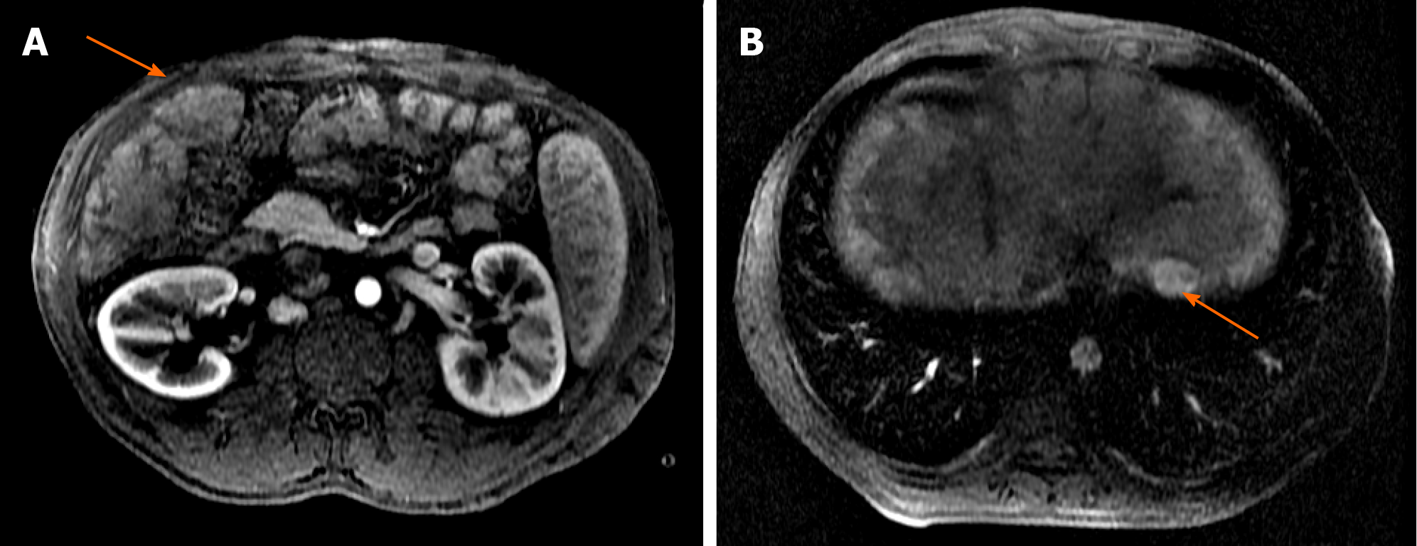
Figure 3 Liver magnetic resonance imaging with hepatobiliary contrast (arterial phase).
A: Hypervascularized nodule in segment V of 4 cm (arrow); B: Hypervascularized nodule in segment II of 2.3 cm (arrow).
- Citation: Rocha-Santos V, Waisberg DR, Pinheiro RS, Nacif LS, Arantes RM, Ducatti L, Martino RB, Haddad LB, Galvao FH, Andraus W, Carneiro-D'Alburquerque LA. Living-donor liver transplantation in Budd-Chiari syndrome with inferior vena cava complete thrombosis: A case report and review of the literature. World J Hepatol 2021; 13(1): 151-161
- URL: https://www.wjgnet.com/1948-5182/full/v13/i1/151.htm
- DOI: https://dx.doi.org/10.4254/wjh.v13.i1.151


