Copyright
©The Author(s) 2020.
World J Hepatol. Apr 27, 2020; 12(4): 160-169
Published online Apr 27, 2020. doi: 10.4254/wjh.v12.i4.160
Published online Apr 27, 2020. doi: 10.4254/wjh.v12.i4.160
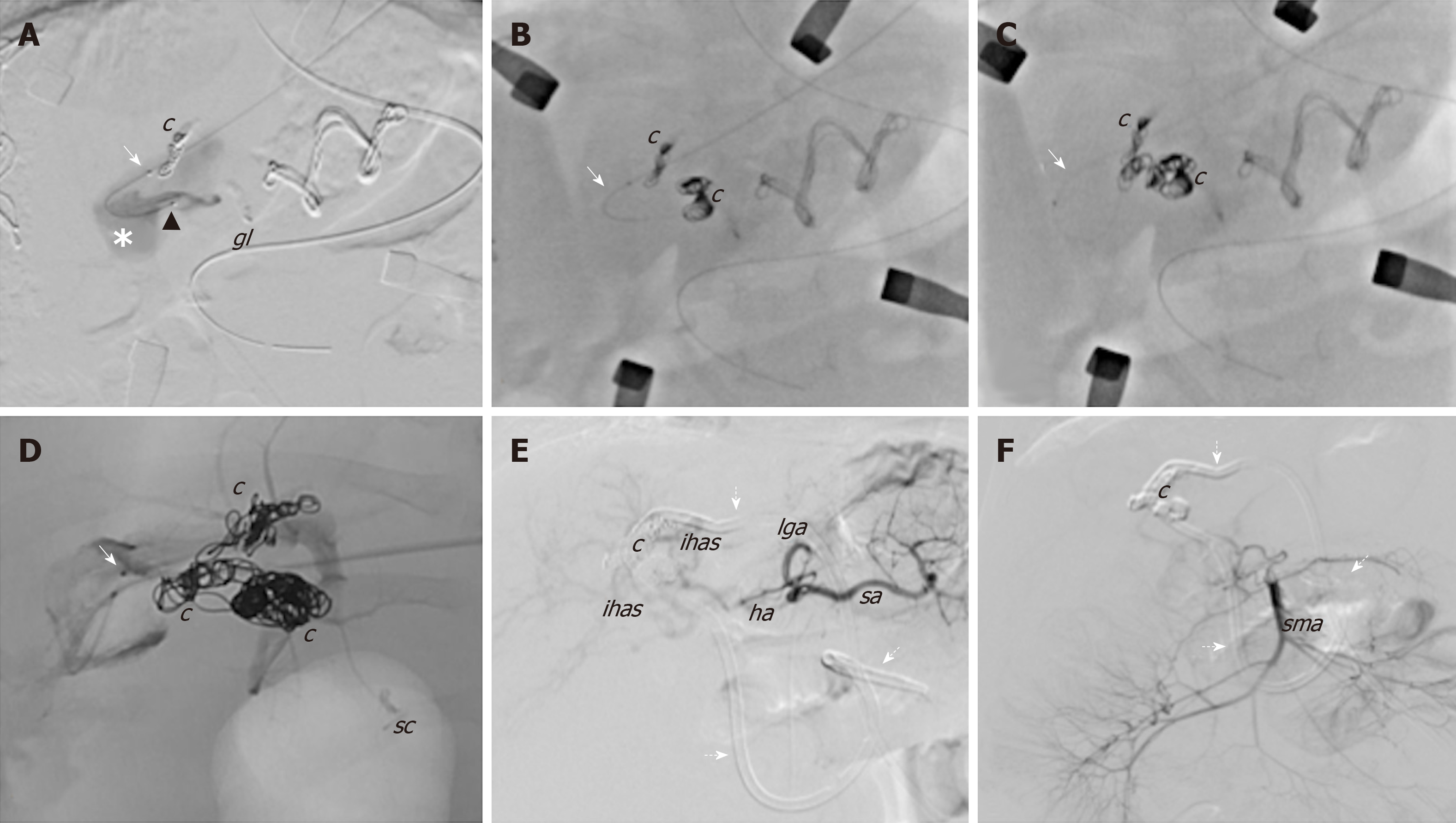
Figure 3 Angiographic images during the hybrid endovascular-surgical procedure of the congenital intrahepatic arterioportal fistula.
A-D: The initial attempts of selective anterograde catheterisation of the vascular malformation failed due to the small size of the arterial branches, and thus a hybrid procedure was performed. It consisted of (1) retrograde venous catheterisation of the fistula through direct left portal vein puncture (white arrow) and embolisation of the shunt with coils; and (2) surgical exposure of the Rex and selective surgical ligation of the small dysplastic arteries feeding the fistula; E and F: Final angiographic images from the celiac trunk and the superior mesenteric artery, which revealed complete closure of the shunt without signs of revascularisation. c: Coils; gl: Glue; ha: Hepatic artery; ihas: Intrahepatic arteries; lga: Left gastric artery; lpa: Left phrenic artery; sma: Superior mesenteric artery; sa: Splenic artery; sc: Surgical clips; Dotted arrows: External internal biliary drainage.
- Citation: Angelico R, Paolantonio G, Paoletti M, Grimaldi C, Saffioti MC, Monti L, Candusso M, Rollo M, Spada M. Combined endovascular-surgical treatment for complex congenital intrahepatic arterioportal fistula: A case report and review of the literature. World J Hepatol 2020; 12(4): 160-169
- URL: https://www.wjgnet.com/1948-5182/full/v12/i4/160.htm
- DOI: https://dx.doi.org/10.4254/wjh.v12.i4.160


