Copyright
©The Author(s) 2020.
World J Hepatol. Apr 27, 2020; 12(4): 160-169
Published online Apr 27, 2020. doi: 10.4254/wjh.v12.i4.160
Published online Apr 27, 2020. doi: 10.4254/wjh.v12.i4.160
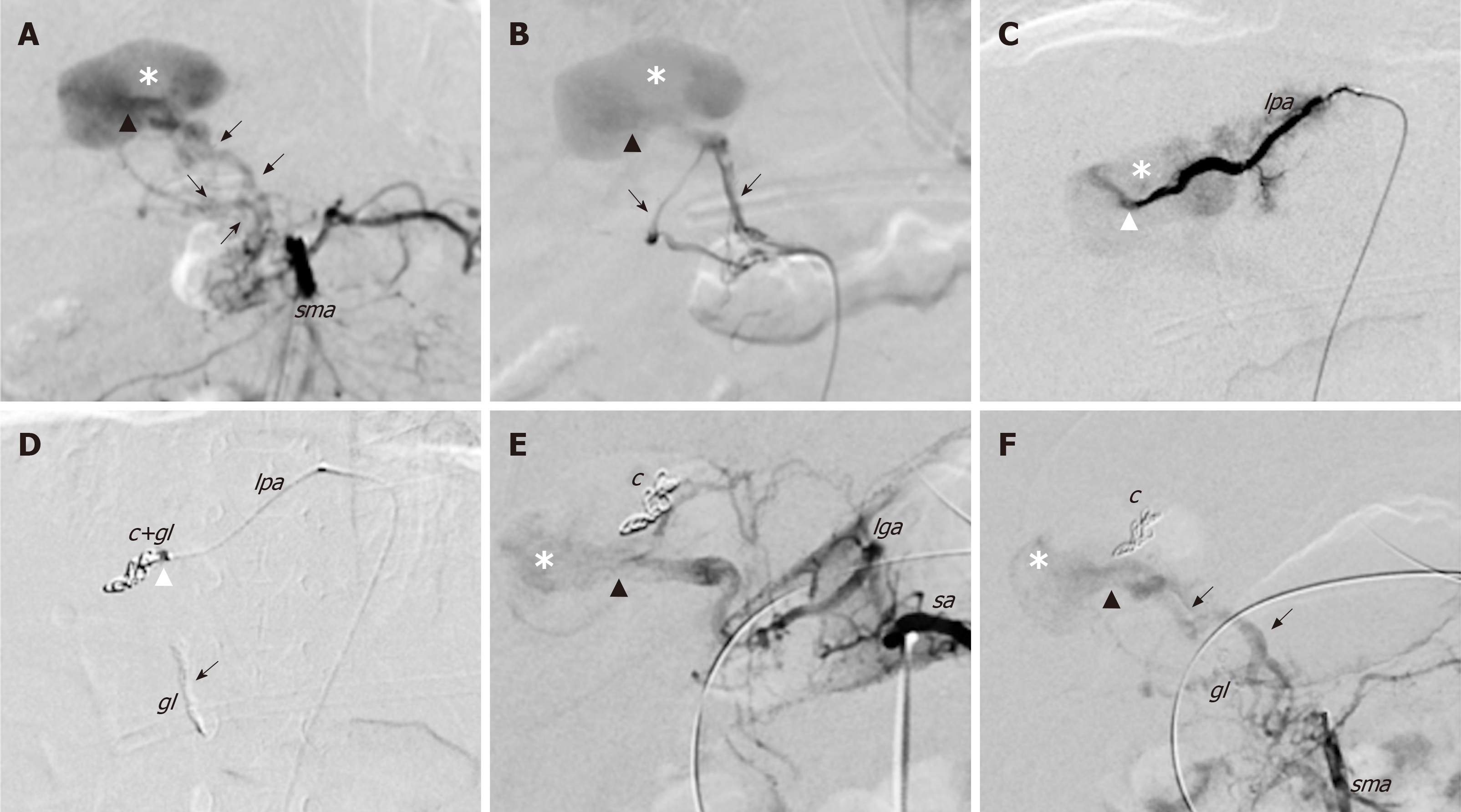
Figure 2 First angiography and endovascular embolization of the congenital intrahepatic arterioportal fistula.
Initial endovascular treatment of the malformation (digital subtraction angiograms). A and B: Angiograms from the superior mesenteric artery show dilated and tortuous dysplastic arteries (black arrows) that converged into the left aneurismal portal vein through one Y-shaped fistula within the Rex recess (black arrow head); C: Superselective catheterisation of a distal branch of the left phrenic artery that shows the additional shunt (white arrow head) into the venous aneurism; D: Embolisation of the shunts with glue and coils with glue cast; E and F: Angiographic control images from celiac trunk (E) and superior mesenteric artery (F) that show persistent patency of the fistula after the embolisation. c: Coils; gl: Glue; lga: Left gastric artery; lpa: Left phrenic artery; sma: Superior mesenteric artery; sa: Splenic artery.
- Citation: Angelico R, Paolantonio G, Paoletti M, Grimaldi C, Saffioti MC, Monti L, Candusso M, Rollo M, Spada M. Combined endovascular-surgical treatment for complex congenital intrahepatic arterioportal fistula: A case report and review of the literature. World J Hepatol 2020; 12(4): 160-169
- URL: https://www.wjgnet.com/1948-5182/full/v12/i4/160.htm
- DOI: https://dx.doi.org/10.4254/wjh.v12.i4.160


