Copyright
©The Author(s) 2019.
World J Hepatol. Mar 27, 2019; 11(3): 318-329
Published online Mar 27, 2019. doi: 10.4254/wjh.v11.i3.318
Published online Mar 27, 2019. doi: 10.4254/wjh.v11.i3.318
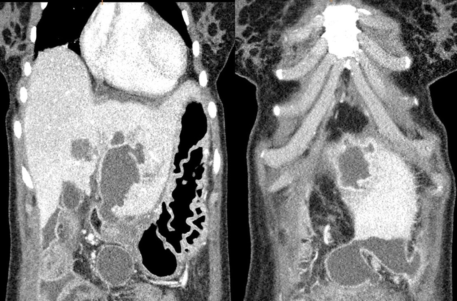
Figure 6 Two different coronal reformatted contrast-enhanced computed tomography images of the same patient show a perforated hydatid cyst located in segment III of the liver and fluid collection in the perihepatic/pelvic area.
- Citation: Akbulut S, Ozdemir F. Intraperitoneal rupture of the hydatid cyst: Four case reports and literature review. World J Hepatol 2019; 11(3): 318-329
- URL: https://www.wjgnet.com/1948-5182/full/v11/i3/318.htm
- DOI: https://dx.doi.org/10.4254/wjh.v11.i3.318


