Copyright
©The Author(s) 2019.
World J Hepatol. Feb 27, 2019; 11(2): 217-225
Published online Feb 27, 2019. doi: 10.4254/wjh.v11.i2.217
Published online Feb 27, 2019. doi: 10.4254/wjh.v11.i2.217
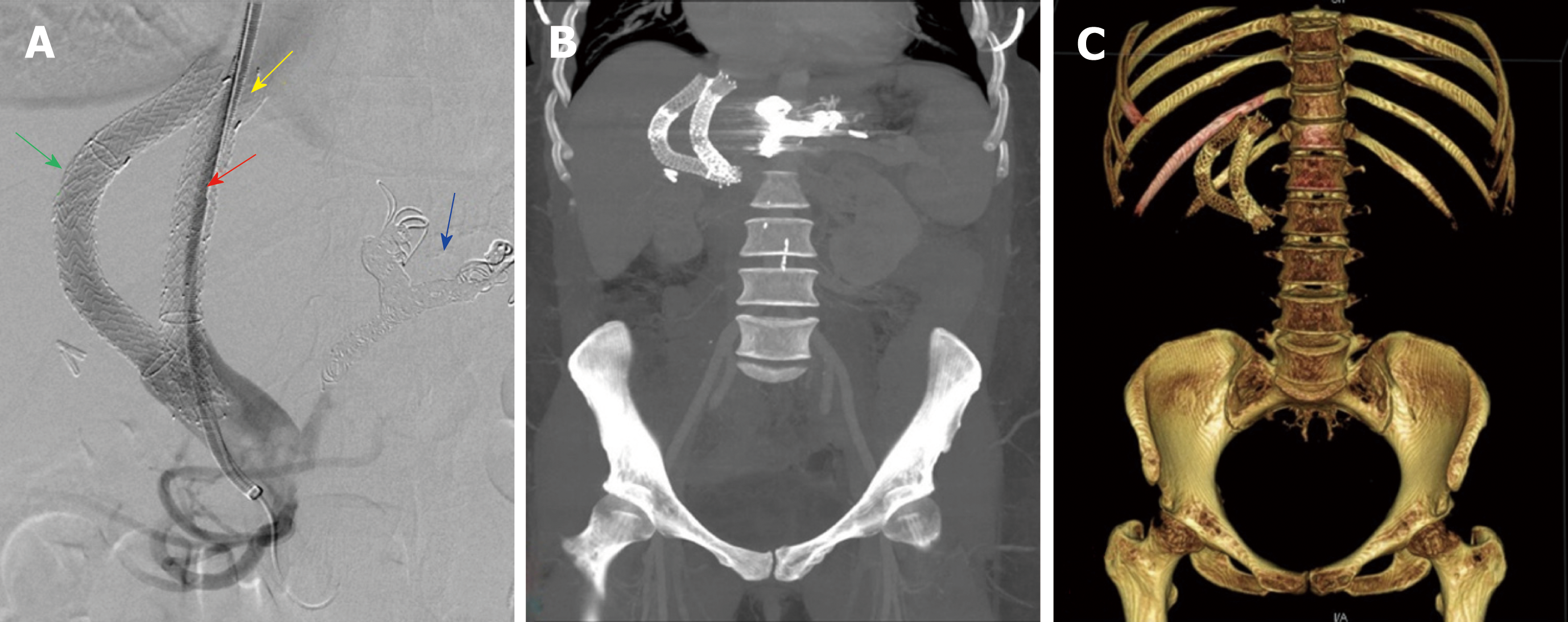
Figure 1 Imaging examinations of patient 1.
A: Digital subtracted portal angiography showing successful placement of the parallel stent (red arrow) with caval extension (yellow arrow). Embolization coils can be seen the coronary vein branches (blue arrow). Primary stent (green arrow) is seen alongside the second transjugular intrahepatic portosystemic shunt stent. B: Maximum intensity projection with 30-mm slab of post procedural computed tomography (CT) abdomen showing primary and parallel stents in tandem. C: 3D reconstruction of a post procedural CT abdomen showing primary and parallel stents in tandem.
- Citation: Raissi D, Yu Q, Nisiewicz M, Krohmer S. Parallel transjugular intrahepatic portosystemic shunt with Viatorr® stents for primary TIPS insufficiency: Case series and review of literature . World J Hepatol 2019; 11(2): 217-225
- URL: https://www.wjgnet.com/1948-5182/full/v11/i2/217.htm
- DOI: https://dx.doi.org/10.4254/wjh.v11.i2.217


