
















©The Author(s) 2019.
World J Hepatol. Dec 27, 2019; 11(12): 773-779
Published online Dec 27, 2019. doi: 10.4254/wjh.v11.i12.773
Published online Dec 27, 2019. doi: 10.4254/wjh.v11.i12.773
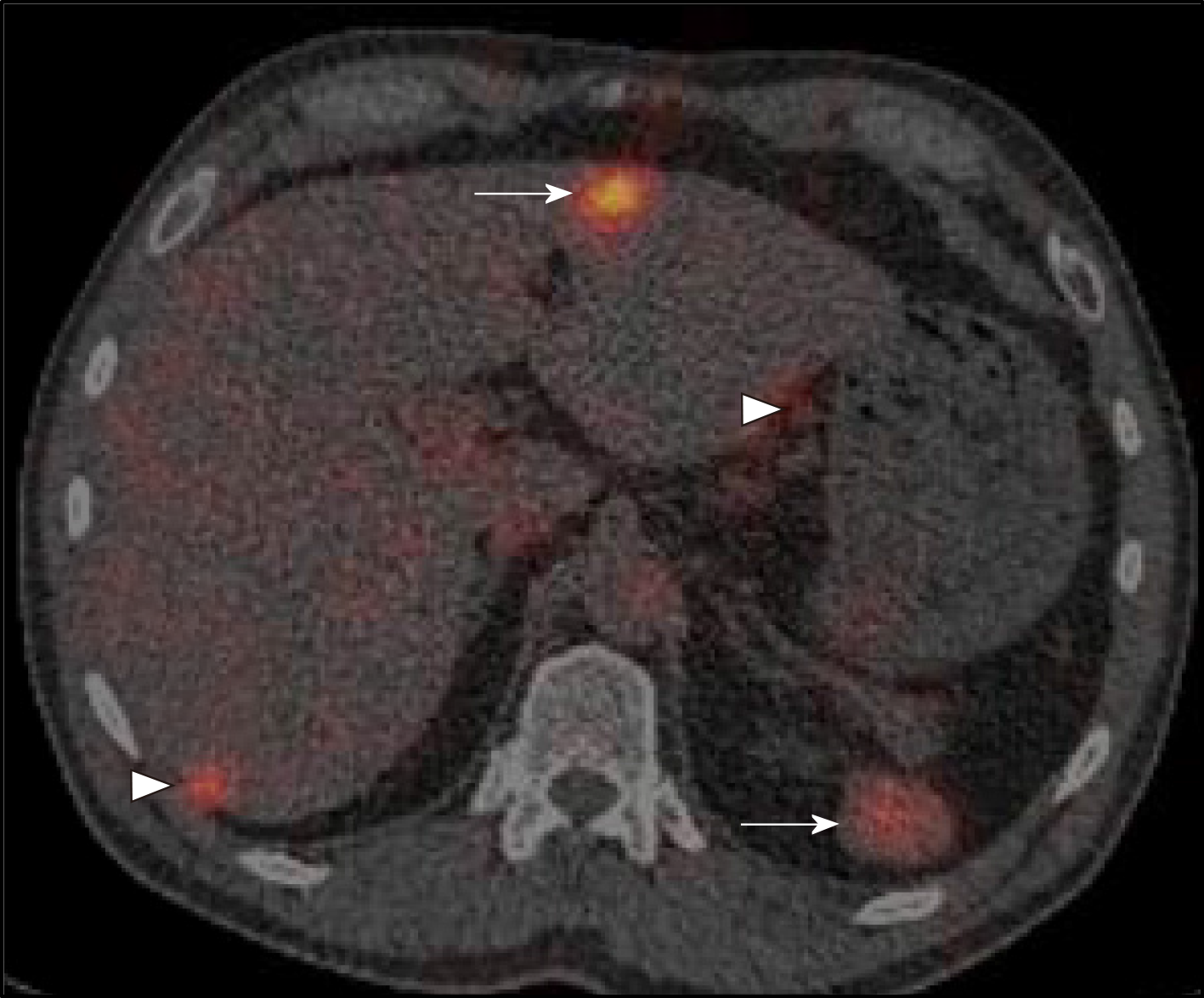
Figure 2 Denatured red cell scan with fused computed tomography images.
The liver lesion (thick arrow), peritoneal and retroperitoneal nodules (arrowheads) and spleen (thin arrow) show uptake in keeping with multiple areas of splenic tissue.
- Citation: Ananthan K, Yusuf GT, Kumar M. Intrahepatic and intra-abdominal splenosis: A case report and review of literature. World J Hepatol 2019; 11(12): 773-779
- URL: https://www.wjgnet.com/1948-5182/full/v11/i12/773.htm
- DOI: https://dx.doi.org/10.4254/wjh.v11.i12.773