Copyright
©The Author(s) 2018.
World J Hepatol. Feb 27, 2018; 10(2): 277-286
Published online Feb 27, 2018. doi: 10.4254/wjh.v10.i2.277
Published online Feb 27, 2018. doi: 10.4254/wjh.v10.i2.277
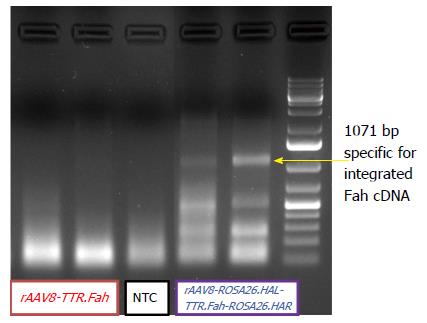
Figure 4 Integration PCR gel electrophoresis.
A representative gel picture from the analyses of genomic liver DNA, that was extracted from snap-frozen liver tissue harvested between 60-70 d after hepatocyte transplantation. Primers were located in the Rosa26 locus and in the FAH sequence of the donor DNA. Product could only be amplified if targeted integration occurred. The expected length of the PCR amplicon was 1107 bp. The PCR product was analysed utilizing agarose gel electrophoresis.
- Citation: Junge N, Yuan Q, Vu TH, Krooss S, Bednarski C, Balakrishnan A, Cathomen T, Manns MP, Baumann U, Sharma AD, Ott M. Homologous recombination mediates stable Fah gene integration and phenotypic correction in tyrosinaemia mouse-model. World J Hepatol 2018; 10(2): 277-286
- URL: https://www.wjgnet.com/1948-5182/full/v10/i2/277.htm
- DOI: https://dx.doi.org/10.4254/wjh.v10.i2.277


