Copyright
©The Author(s) 2018.
World J Hepatol. Oct 27, 2018; 10(10): 772-779
Published online Oct 27, 2018. doi: 10.4254/wjh.v10.i10.772
Published online Oct 27, 2018. doi: 10.4254/wjh.v10.i10.772
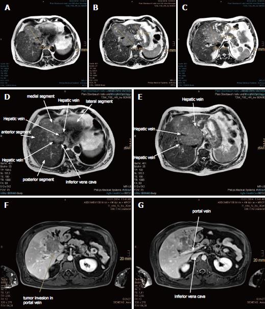
Figure 1 Large hepatocellular carcinoma with vascular invasion and satellite metastases in the magnetic resonance imaging scan prior to proton beam therapy.
A: The upper portion of the main tumor abutting the heart in axial plane is shown; B: Extended hepatocellular carcinoma (HCC) with invasion of portal and hepatic veins in the axial plane is shown; C: The lower portion of the main tumor abutting the inferior vena cava is shown, with dilated segmental bile ducts and 2 liver metastases in the left hepatic lobe; D-G: The HCC involved all three hepatic veins as well as the portal vein. The inferior vena cava was also compressed.
- Citation: Lin YL. Proton beam therapy in apneic oxygenation treatment of an unresectable hepatocellular carcinoma: A case report and review of literature. World J Hepatol 2018; 10(10): 772-779
- URL: https://www.wjgnet.com/1948-5182/full/v10/i10/772.htm
- DOI: https://dx.doi.org/10.4254/wjh.v10.i10.772


