Copyright
©The Author(s) 2018.
World J Hepatol. Jan 27, 2018; 10(1): 22-33
Published online Jan 27, 2018. doi: 10.4254/wjh.v10.i1.22
Published online Jan 27, 2018. doi: 10.4254/wjh.v10.i1.22
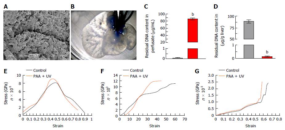
Figure 3 Characterization of acellularized liver scaffold.
A: Ultra-structural characterization of acellularized xenogenic liver showing acellularity in the scaffold with preserved three-dimensional microanatomy of the portal tract surrounded by lobular structures. The parenchymal spaces were found enriched with connective tissue fibres arranged with honeycomb-like structures which are an exceptionally preserved anatomy of connective tissues which structures the hepatocyte-free spaces; B: Methylene blue dye infusion showing intact vasculature post-acellularization; Residual DNA content in liver perfusate (C) and in liver tissues (D) before (black bar) and after acellularization (red bar) (bP < 0.0001); E: Mixed tensile strength; F: Mixed suture retention strength; G: Mixed comprehensive strength plots showing higher degree of retention of mechanical properties post-acellularization.
- Citation: Vishwakarma SK, Bardia A, Lakkireddy C, Nagarapu R, Habeeb MA, Khan AA. Bioengineered humanized livers as better three-dimensional drug testing model system. World J Hepatol 2018; 10(1): 22-33
- URL: https://www.wjgnet.com/1948-5182/full/v10/i1/22.htm
- DOI: https://dx.doi.org/10.4254/wjh.v10.i1.22


