Copyright
©2010 Baishideng Publishing Group Co.
World J Hepatol. Aug 27, 2010; 2(8): 318-321
Published online Aug 27, 2010. doi: 10.4254/wjh.v2.i8.318
Published online Aug 27, 2010. doi: 10.4254/wjh.v2.i8.318
Figure 1 Digital subtraction angiography.
A: Digital subtraction angiography showed tumor staining close against the diaphragm 20 mm in diameter; B: Computed tomography (CT) during arterial portography showed a perfusion defect in segment 8; C: CT during hepatic arteriography showed a hypervascularity lesion in the corresponding region.
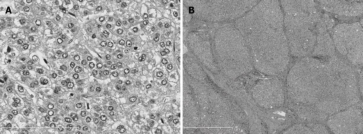
Figure 2 Histological findings of specimens obtained by subsegmentectomy.
A: The tumor is a moderately differentiated hepatocellular carcinoma (H&E stain, × 100). B: The non-tumorous liver tissue around the tumor shows cirrhosis (H&E stain, × 40).
- Citation: Akima T, Tamano M, Yamagishi H, Kubota K, Fujimori T, Hiraishi H. Surgical treatment of HCC in a patient with lamivudine-resistant hepatitis B cirrhosis with adefovir dipivoxil. World J Hepatol 2010; 2(8): 318-321
- URL: https://www.wjgnet.com/1948-5182/full/v2/i8/318.htm
- DOI: https://dx.doi.org/10.4254/wjh.v2.i8.318


