Copyright
©2010 Baishideng Publishing Group Co.
World J Hepatol. Oct 27, 2010; 2(10): 387-391
Published online Oct 27, 2010. doi: 10.4254/wjh.v2.i10.387
Published online Oct 27, 2010. doi: 10.4254/wjh.v2.i10.387
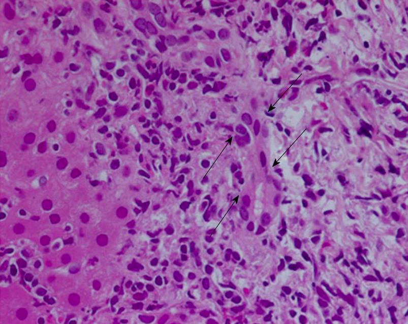
Figure 1 Histopathological findings of the liver biopsy.
Periportal lymphocytic infiltration with bile duct damage (arrows: damaged bile duct; hematoxylin and eosin stain, × 400).
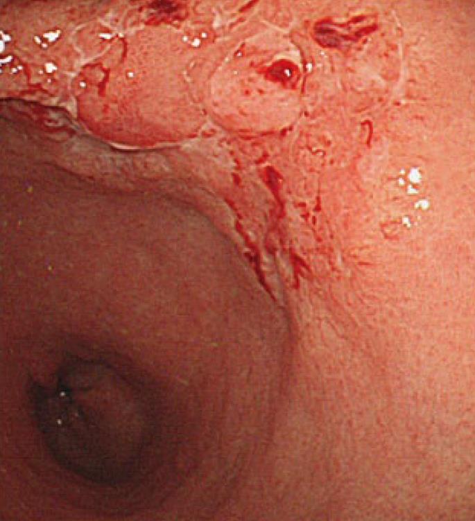
Figure 2 Endoscopic picture of the stomach.
The depressed lesion with spontaneous hemorrhage and irregular margins.
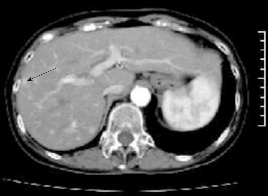
Figure 3 Contrast-enhanced abdominal computed tomography.
A well-circumscribed nodule showing early arterial enhancement in S8 (arrow).
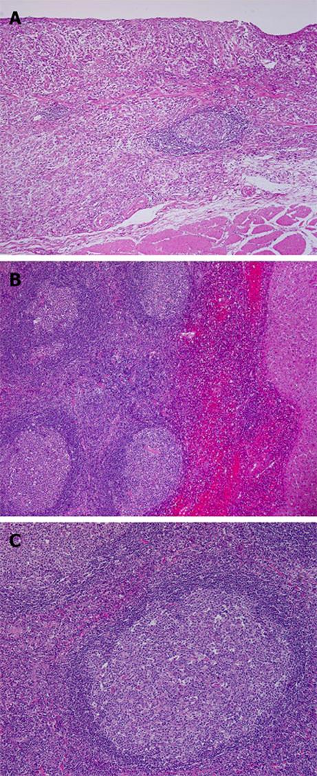
Figure 4 Histopathological findings of the gastric carcinoma and liver nodule.
A: Signet ring cell carcinoma of the stomach invading into the deep submucosa (hematoxylin and eosin stain, × 40); B: A relatively well-circumscribed nodular proliferation of lymphocytes with lymphoid follicles in the liver (right: liver parenchyma, hematoxylin and eosin stain, × 40); C: Mantle zone and germinal center are sharply demarcated and a mixture of small and large lymphocytes and scattered tingible body macrophages is observed in the germinal center (hematoxylin and eosin stain, × 40).
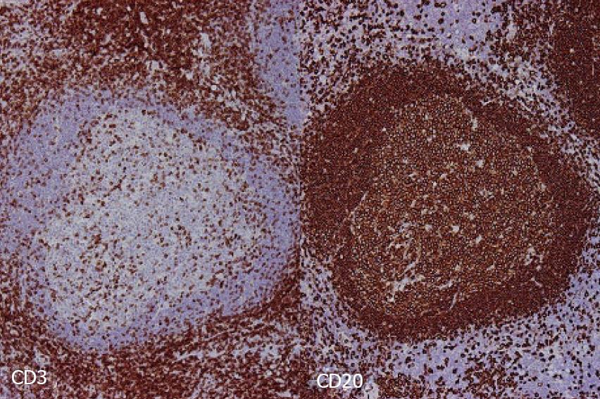
Figure 5 Immunohistochemical findings of the liver nodule.
CD3-positive T lymphocytes (left) and CD20-positive B lymphocytes (right) are regularly distributed (× 40).
- Citation: Ishida M, Nakahara T, Mochizuki Y, Tsujikawa T, Andoh A, Saito Y, Yamamoto H, Kojima F, Hotta M, Tani T, Fujiyama Y, Okabe H. Hepatic reactive lymphoid hyperplasia in a patient with primary biliary cirrhosis. World J Hepatol 2010; 2(10): 387-391
- URL: https://www.wjgnet.com/1948-5182/full/v2/i10/387.htm
- DOI: https://dx.doi.org/10.4254/wjh.v2.i10.387


