Copyright
©The Author(s) 2023.
World J Hepatol. Jan 27, 2023; 15(1): 52-67
Published online Jan 27, 2023. doi: 10.4254/wjh.v15.i1.52
Published online Jan 27, 2023. doi: 10.4254/wjh.v15.i1.52
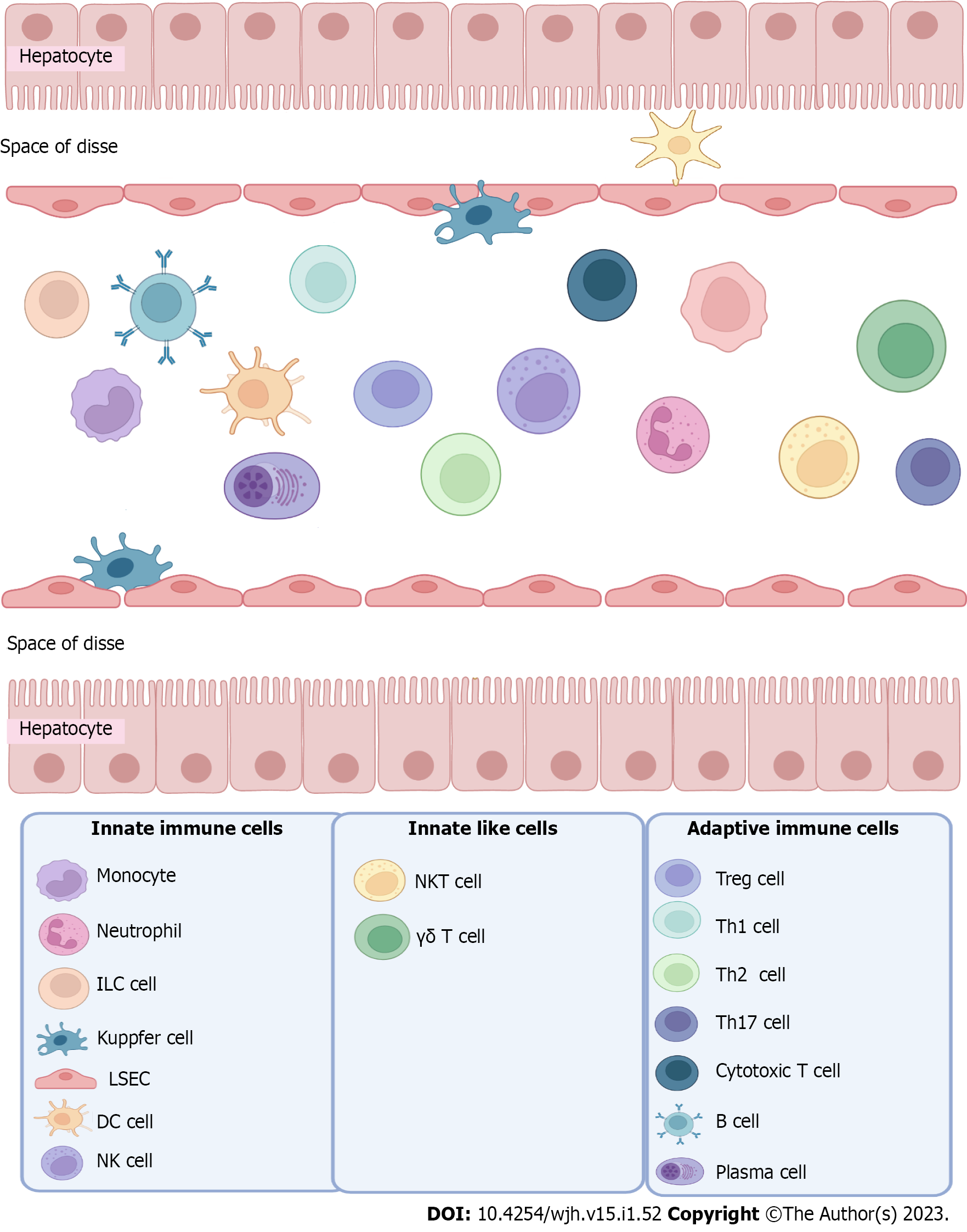
Figure 1 Cell composition of the healthy liver.
ILC: Innate lymphoid cells; DC: Dendritic cells; LSEC: Liver sinusoidal endothelial cells; NK: Natural killer; NKT: Natural killer T.
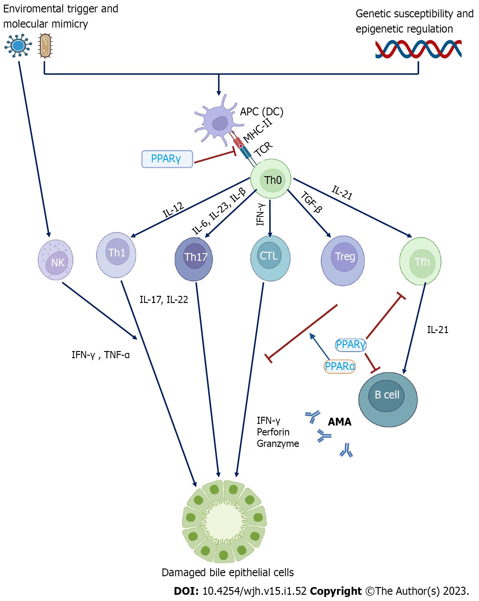
Figure 2 Model of pathogenic mechanisms in primary biliary cholangitis.
APC: Antigen presenting cell; DC: Dendritic cell; NK: Natural killer; IL: Interleukin; TNF: Tumor necrosis factor; TGF-β: Transforming growth factor β; IFN-γ: Interferon-gamma; PPAR: Peroxisome proliferator-activated receptor; AMA: Anti-mitochondrial antibody.
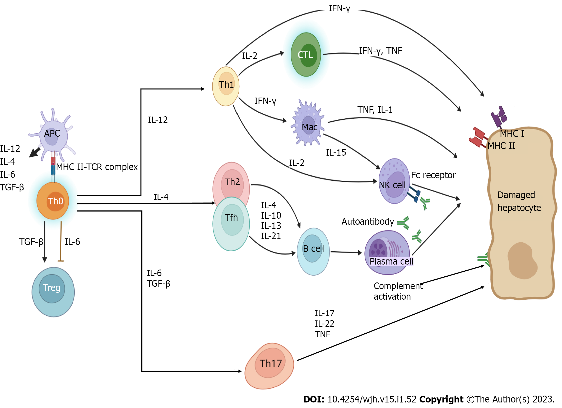
Figure 3 Pathogenic pathways of autoimmune hepatitis.
APC: Antigen-presenting cell; CTL: Cytotoxic T lymphocyte; Mac: Macrophage; IL: Interleukin; TNF: Tumor necrosis factor; MHC: Major histocompatibility complex; TGF-β: Transforming growth factor β; IFN-γ: Interferon-gamma.
- Citation: Parlar YE, Ayar SN, Cagdas D, Balaban YH. Liver immunity, autoimmunity, and inborn errors of immunity. World J Hepatol 2023; 15(1): 52-67
- URL: https://www.wjgnet.com/1948-5182/full/v15/i1/52.htm
- DOI: https://dx.doi.org/10.4254/wjh.v15.i1.52


