Copyright
©The Author(s) 2015.
World J Stem Cells. Sep 26, 2015; 7(8): 1090-1108
Published online Sep 26, 2015. doi: 10.4252/wjsc.v7.i8.1090
Published online Sep 26, 2015. doi: 10.4252/wjsc.v7.i8.1090
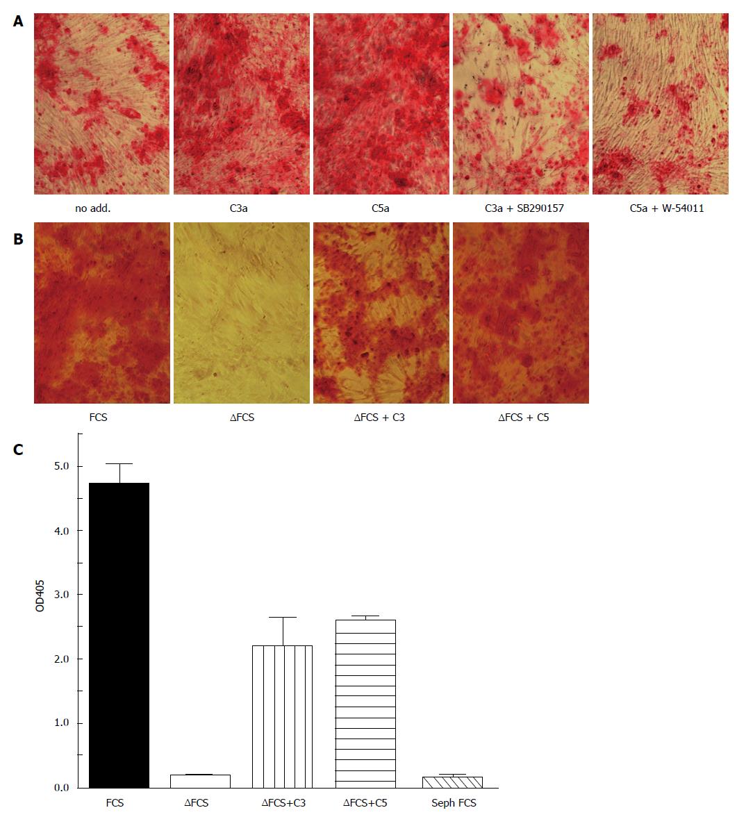
Figure 2 Complement activation accelerates osteogenic differentiation of bone marrow mesenchymal stem cells.
MSC were cultured in osteogenic media (α-MEM containing 16.5% FCS, not heat inactivated, 10 nmol/L dexamethasone, 20 mmol/L β-glycerolphosphate, and 50 μmol/L ascorbic acid 2-phosphate) for the indicated time. Osteogenesis was detected by alizarin red S staining. A: All cells were cultured in the presence of the carboxypeptidase inhibitor 2-mercaptomethyl-3-guanidinoethylthioproprionic acid (80 nmol/L) to maintain C3a and C5a activity. C3a (100 nmol/L) or C5a (10 nmol/L) were added during the first 3 d of cultures in the presence or absence of the C3aR inhibitor SB290157 (1 nmol/L) or the C5aR inhibitor W-54001 (1 nmol/L). Both C3a and C5a accelerated calcification in a C3aR and C5aR specific fashion as detected by Alizarin red staining on day 14; B: Osteogenesis was considerably delayed in heat-inactivated FCS (FCS), in which complement components are inactivated. Addition of either C3 or C5 partially reconstituted the effect of FCS. Alizarin staining on day 21; C: Quantitation of the alizarin staining of figure 2B following solubilizing in acid SDS solution. FCS: Fetal calf serum; MSC: Mesenchymal stem cells.
- Citation: Schraufstatter IU, Khaldoyanidi SK, DiScipio RG. Complement activation in the context of stem cells and tissue repair. World J Stem Cells 2015; 7(8): 1090-1108
- URL: https://www.wjgnet.com/1948-0210/full/v7/i8/1090.htm
- DOI: https://dx.doi.org/10.4252/wjsc.v7.i8.1090


