Copyright
©The Author(s) 2015.
World J Stem Cells. Aug 26, 2015; 7(7): 1064-1077
Published online Aug 26, 2015. doi: 10.4252/wjsc.v7.i7.1064
Published online Aug 26, 2015. doi: 10.4252/wjsc.v7.i7.1064
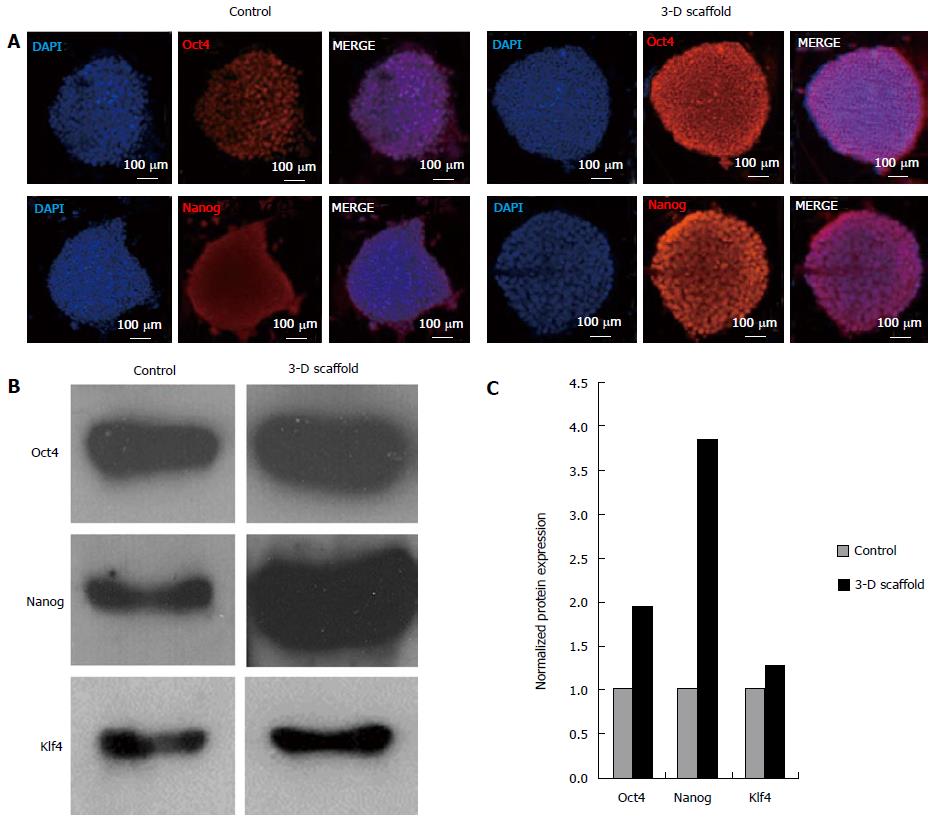
Figure 4 Protein expression of pluripotency markers in embryonic stem cells grown in two-dimensional and three-dimensional cultures.
ESCs grown in two-dimensional (2-D) and in 3-D floating scaffolds (prepared with 33% thiol substitution) were analyzed by immunofluorescence staining and western blotting. A: Cells grown in 2-D culture and 3-D scaffolds for 2 wk were cultured on coverslips for 2 d and stained with primary antibodies, Oct4 and Nanog, treated with Alexa Fluor 568 conjugated secondary antibodies, and counterstained with DAPI. Confocal images depicted an increase in Oct4 and Nanog protein expression in 3-D scaffold grown ESCs compared to 2-D cultured cells; B: Western blot analysis showed increased expressions of Oct4, Nanog, and Klf4 proteins in ESCs grown in the 3-D scaffold for 2 wk compared to initial 2-D grown ESCs (day 0). Cell were lysed in RIPA buffer and aliquots containing an equal amount of protein (10 μg) were subjected to 12% polyacrylamide gel electrophoresis, transferred to nitrocellulose membranes, and probed with antibodies against Oct4, Nanog, and Klf4; C: Quantitative analysis of western blots (normalized to β-actin levels) showed that Oct4, Nanog and Klf4 protein levels in 3-D grown cells were increased by 1.9, 3.9 and 1.3 fold, respectively, as compared to 2-D cultured controls. Representative results are shown. ESCs: Embryonic stem cells. DAPI: 4′,6-diamidino-2-phenylindole dihydrochloride.
- Citation: McKee C, Perez-Cruet M, Chavez F, Chaudhry GR. Simplified three-dimensional culture system for long-term expansion of embryonic stem cells. World J Stem Cells 2015; 7(7): 1064-1077
- URL: https://www.wjgnet.com/1948-0210/full/v7/i7/1064.htm
- DOI: https://dx.doi.org/10.4252/wjsc.v7.i7.1064


