Copyright
©The Author(s) 2015.
World J Stem Cells. Mar 26, 2015; 7(2): 521-534
Published online Mar 26, 2015. doi: 10.4252/wjsc.v7.i2.521
Published online Mar 26, 2015. doi: 10.4252/wjsc.v7.i2.521
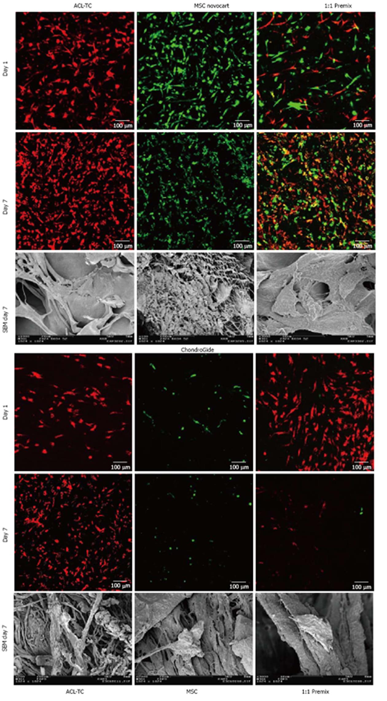
Figure 4 Z-projections of confocal laser scanning microscope images of 200 μm 3D-stacks through the collagen patches of mesenchymal stem cells and anterior cruciate ligament-derived tenocytes on day 1 and after 7 d of culture on Novocart™ and Chondro-Gide™.
Cells were stained with Vybrant™ DiO (DiO = green, MSCs) and DiL membrane tracker dyes (DiL = red, TCs) prior to seeding. Last row are scanning electron microscope pictures of the cell-seeded scaffolds after 7 d of culture showing adherence and shape of the MSCs and tenocytes on the scaffolds. Note that for Chondro-Gide™ patches these are only images from TCs seeded scaffolds. For the Novocart™ patches the right most image is from an experiment containing a premix of cells, and the left most and center SEM image are from experiments with MSCs seeded scaffolds. MSC: Mesenchymal stem cell; ACL-TC: Anterior cruciate ligament-derived tenocyte; SEM: Scanning electron microscope.
- Citation: Gantenbein B, Gadhari N, Chan SC, Kohl S, Ahmad SS. Mesenchymal stem cells and collagen patches for anterior cruciate ligament repair. World J Stem Cells 2015; 7(2): 521-534
- URL: https://www.wjgnet.com/1948-0210/full/v7/i2/521.htm
- DOI: https://dx.doi.org/10.4252/wjsc.v7.i2.521


