Copyright
©2013 Baishideng Publishing Group Co.
World J Stem Cells. Jul 26, 2013; 5(3): 86-97
Published online Jul 26, 2013. doi: 10.4252/wjsc.v5.i3.86
Published online Jul 26, 2013. doi: 10.4252/wjsc.v5.i3.86
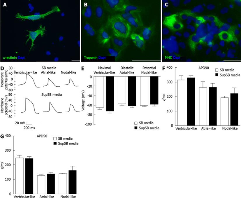
Figure 8 Characterization of cardiomyocytes: Immunocytochemistry and electrophysiology.
HES-3 differentiated cardiomyocyte aggregates at the end of differentiation (day 16) were mechanically dissociated and plated on 24-well plates and strained for markers. A: Sarcomeric α-actinin (Structural protein); B: Troponin-T (contractile function protein); C: Myosin heavy chain (MHC) (contractile function protein). Nuclei stained with DAPI (blue). Scale bar = 20 μm. MHC, myosin heavy chain; DAPI: 4’6-diamidino-2-phenylindole. D: Whole-cell patch-clamp recording was performed on beating cardiomyocyte aggregates at day 23 of differentiation. The recording shows the successful derivation of all three cardiac phenotypes as well as no difference between cells grown in SB media (control) and SupSB media (supplements); E: Maximal diastolic potential; F: Action potential duration at 90% repolarization (ADP90); G: Action potential duration at 50% repolarization (ADP50).
- Citation: Ting S, Lecina M, Chan YC, Tse HF, Reuveny S, Oh SK. Nutrient supplemented serum-free medium increases cardiomyogenesis efficiency of human pluripotent stem cells. World J Stem Cells 2013; 5(3): 86-97
- URL: https://www.wjgnet.com/1948-0210/full/v5/i3/86.htm
- DOI: https://dx.doi.org/10.4252/wjsc.v5.i3.86


