Copyright
©2012 Baishideng.
World J Stem Cells. Jun 26, 2012; 4(6): 53-61
Published online Jun 26, 2012. doi: 10.4252/wjsc.v4.i6.53
Published online Jun 26, 2012. doi: 10.4252/wjsc.v4.i6.53
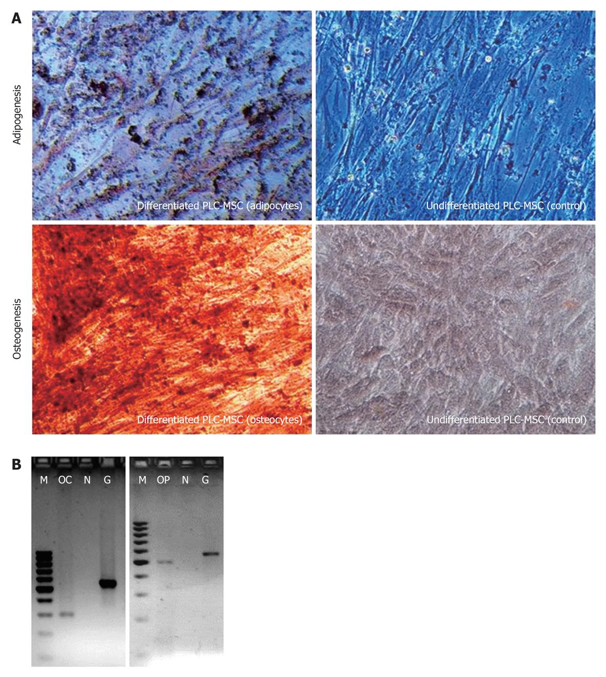
Figure 4 Differentiation potential of placenta mesenchymal stem cells into mesodermal lineages.
A: Placenta MSC (PLC-MSC) after 3 wk in adipogenic or osteogenic or normal cell culture medium. Formation of lipid droplets (stained red in Oil-Red-O), calcium deposition (stained orangy-red in Alizarin Red) and formation of proteoglycans (stained blue in Alcian Blue) confirms the ability to differentiate into mesodermal lineages. The picture was taken using phase contrast microscope at 100 × magnification. B: PLC-MSC differentiated into osteocytes expresses Osteocalcin (OC) and Osteopontin (OP) (data from two independent experiments). M: 100 bp DNA ladder; N: Non template control as negative control; C: Control-undifferentiated PLC-MSC; G1: GAPDH for differentiated PLC-MSC; G2: GAPDH for control.
- Citation: Vellasamy S, Sandrasaigaran P, Vidyadaran S, George E, Ramasamy R. Isolation and characterisation of mesenchymal stem cells derived from human placenta tissue. World J Stem Cells 2012; 4(6): 53-61
- URL: https://www.wjgnet.com/1948-0210/full/v4/i6/53.htm
- DOI: https://dx.doi.org/10.4252/wjsc.v4.i6.53


