Copyright
©2012 Baishideng.
World J Stem Cells. Apr 26, 2012; 4(4): 21-27
Published online Apr 26, 2012. doi: 10.4252/wjsc.v4.i4.21
Published online Apr 26, 2012. doi: 10.4252/wjsc.v4.i4.21
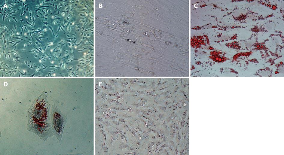
Figure 1 Human umbilical mesenchymal stem cells, adipogenic differentiation and red-o staining.
A is the human mesenchymal stem cells at passage 4, the cells assumed a more spindle-shaped, fibroblastic morphology; B-D demonstrate the human umbilical mesenchymal stem cells (HUMSCs) after 2 wk adipogenic differentiation without and with the Red-O staining, respectively. The differentiated cells’ shape had transformed into an ovoid/round morphology and there were abundant lipid droplets presented in the cell periphery; E is the HUMSCs with Red-O staining without differentiation, no lipid body is visible.
- Citation: Xu ZF, Pan AZ, Yong F, Shen CY, Chen YW, Wu RH. Human umbilical mesenchymal stem cell and its adipogenic differentiation: Profiling by nuclear magnetic resonance spectroscopy. World J Stem Cells 2012; 4(4): 21-27
- URL: https://www.wjgnet.com/1948-0210/full/v4/i4/21.htm
- DOI: https://dx.doi.org/10.4252/wjsc.v4.i4.21


