Copyright
©2011 Baishideng Publishing Group Co.
World J Stem Cells. Jun 26, 2011; 3(6): 53-62
Published online Jun 26, 2011. doi: 10.4252/wjsc.v3.i6.53
Published online Jun 26, 2011. doi: 10.4252/wjsc.v3.i6.53
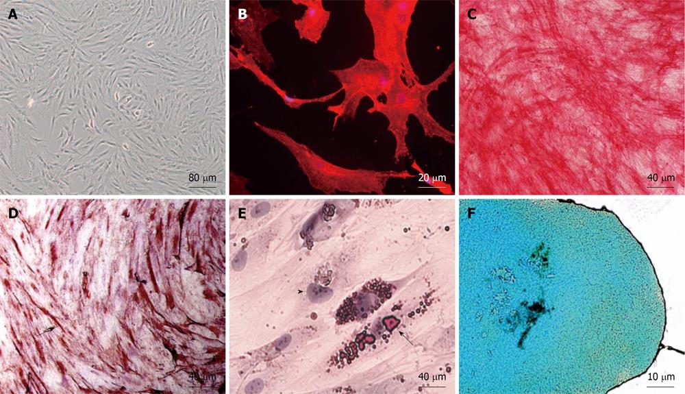
Figure 1 In vitro characterization of human adipose-derived mesenchymal stem cell.
A: Phase contrast of a primary subconfluent culture showing typical adherent fibroblast-like morphology; B: Anti-CD90 immunofluorescence (in red) of a human adipose-derived mesenchymal stem cell (hAMSC) culture; C: Assessment of in vitro differentiation potential of hAMSC by osteogenic differentiation, demonstrated by alkaline phosphatase activity in red; D: Assessment of in vitro differentiation potential of hAMSC by myogenic differentiation, demonstrated by anti-sarcomeric-α-actin in brown (arrow); E: Assessment of in vitro differentiation potential of hAMSC by adipogenic differentiation, demonstrated by Oil red. Arrow: fat drops in a labeled differentiated cell. Arrowhead: Nucleus of an undifferentiated cell; F: Assessment of in vitro differentiation potential of hAMSC by chondrogenic differentiation, demonstrated by Alcian blue.
- Citation: López-Iglesias P, Blázquez-Martínez A, Fernández-Delgado J, Regadera J, Nistal M, Miguel MPD. Short and long term fate of human AMSC subcutaneously injected in mice. World J Stem Cells 2011; 3(6): 53-62
- URL: https://www.wjgnet.com/1948-0210/full/v3/i6/53.htm
- DOI: https://dx.doi.org/10.4252/wjsc.v3.i6.53


