Copyright
©2011 Baishideng Publishing Group Co.
World J Stem Cells. Apr 26, 2011; 3(4): 34-42
Published online Apr 26, 2011. doi: 10.4252/wjsc.v3.i4.34
Published online Apr 26, 2011. doi: 10.4252/wjsc.v3.i4.34
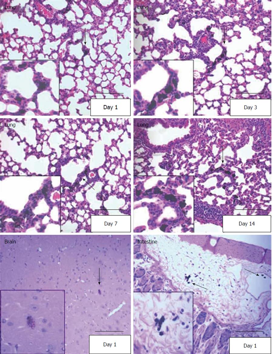
Figure 6 Visualization of human umbilical cord matrix stem cells in hematoxylin and eosin stained tissue sections.
Human umbilical cord matrix stem (hUCMS) cells were loaded with India Black ink and injected through the tail vein. hUCMS cells were detected in lung, intestine and brain on the days indicated in the panels at 200 × magnification. Red arrows in the 200 × pictures indicate India Black ink-labeled hUCMS cells which were magnified at 400 × magnification and presented as inset in each panel. Scale bar = 50 μm.
- Citation: Maurya DK, Doi C, Pyle M, Rachakatla RS, Davis D, Tamura M, Troyer D. Non-random tissue distribution of human naïve umbilical cord matrix stem cells. World J Stem Cells 2011; 3(4): 34-42
- URL: https://www.wjgnet.com/1948-0210/full/v3/i4/34.htm
- DOI: https://dx.doi.org/10.4252/wjsc.v3.i4.34


