
















Copyright: ©Author(s) 2026.
World J Stem Cells. Aug 26, 2026; 18(8): 120270
Published online Aug 26, 2026. doi: 10.4252/wjsc.120270
Published online Aug 26, 2026. doi: 10.4252/wjsc.120270
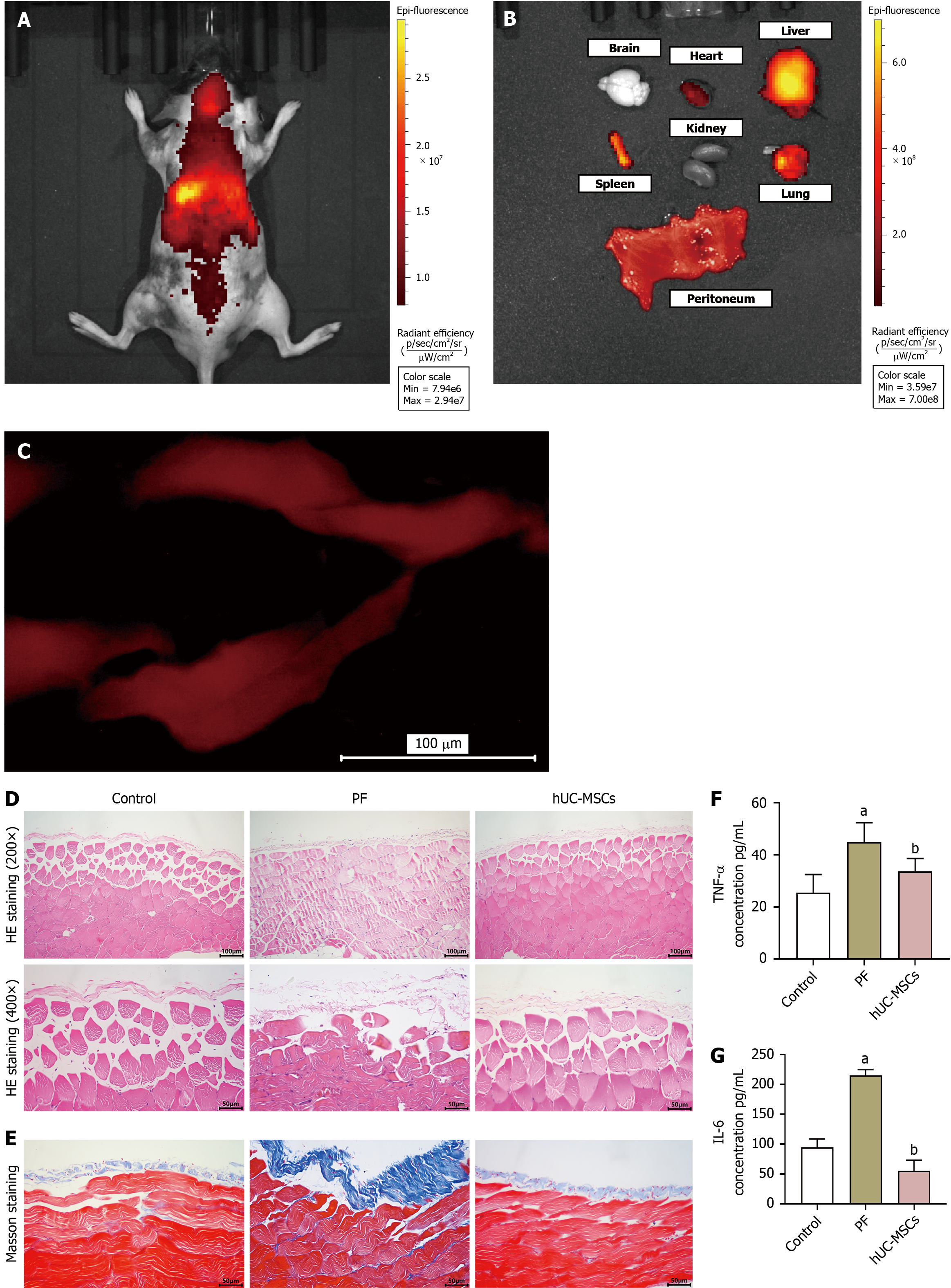
Figure 6 Effects of human umbilical cord-derived mesenchymal stem cells on peritoneal fibrosis and inflammatory cytokines in peritoneal fibrosis mice.
A-C: Tracking the distribution of human umbilical cord-derived mesenchymal stem cells (hUC-MSCs) injected in mice. In vivo imaging of whole-body fluorescence (A). Fluorescent imaging of isolated organs (B). Fluorescence imaging of peritoneal tissues following hUC-MSC injection (C); D: Representative hematoxylin and eosin staining images; E: Representative Masson staining images; F and G: Tumor necrosis factor-alpha (F) and interleukin-6 (G) levels in peritoneal tissues were detected via enzyme-linked immunosorbent assay. Data are presented as mean ± SD. aP < 0.05 vs control; bP < 0.05 vs peritoneal fibrosis. hUC-MSCs: Human umbilical cord-derived mesenchymal stem cells; PF: Peritoneal fibrosis; HE: Hematoxylin and eosin; TNF-α: Tumor necrosis factor-alpha; IL-6: Interleukin-6.
- Citation: Dong FX, Zheng LL, Nie K, Wan JX. Protective effect of hUC-MSCs regulating peritoneal mesothelial cell EMT through cGAS-STING signaling pathway in high glucose-induced peritoneal fibrosis. World J Stem Cells 2026; 18(8): 120270
- URL: https://www.wjgnet.com/1948-0210/full/v18/i8/120270.htm
- DOI: https://dx.doi.org/10.4252/wjsc.120270