















Copyright: ©Author(s) 2026.
World J Stem Cells. Jul 26, 2026; 18(7): 120578
Published online Jul 26, 2026. doi: 10.4252/wjsc.120578
Published online Jul 26, 2026. doi: 10.4252/wjsc.120578
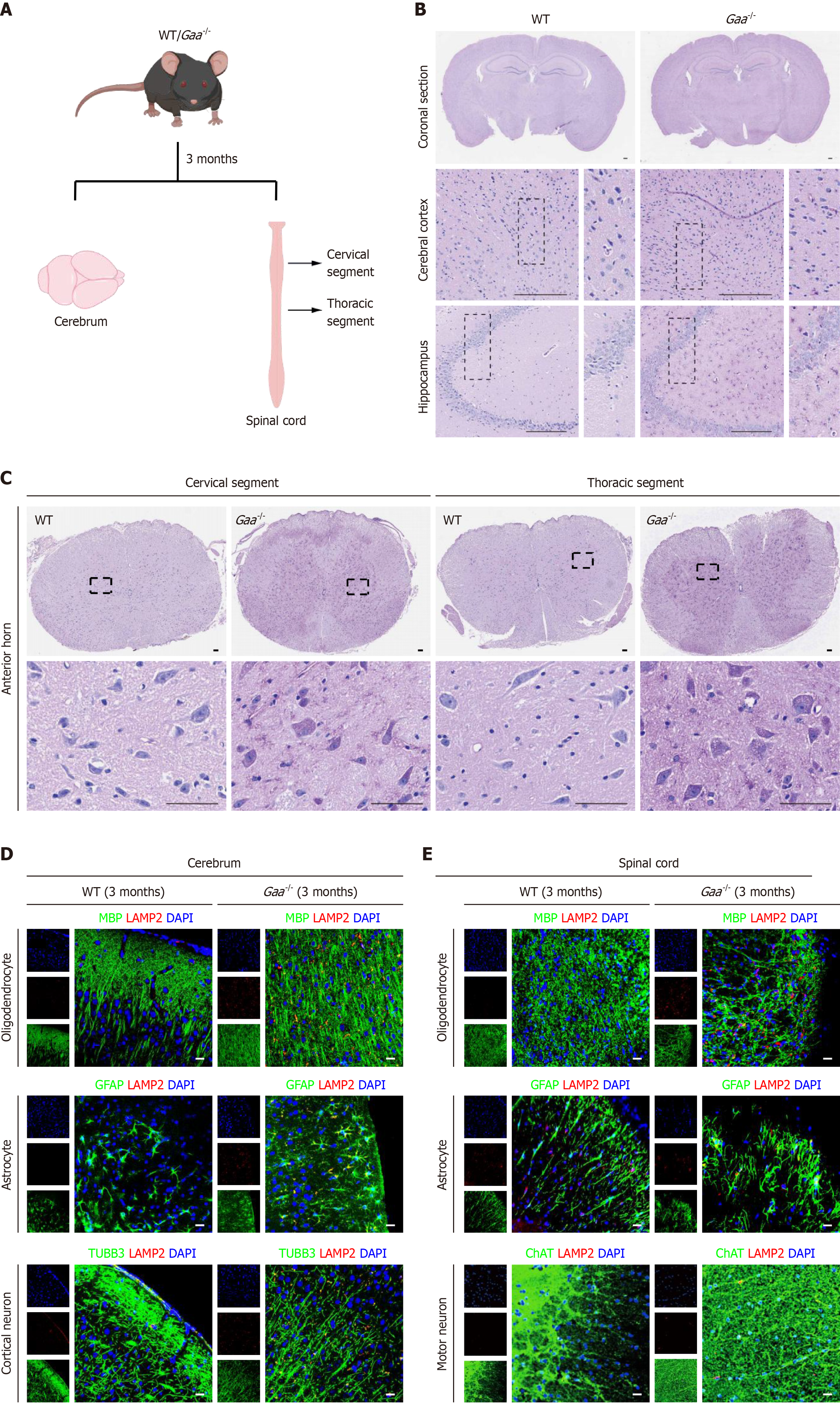
Figure 3 Central nervous system lysosomal pathology in Gaa-/- mice.
A: Schematic overview of tissue sampling from the brain and spinal cord of 3-month-old Gaa-/- mice; B: PAS staining reveals glycogen accumulation (magenta granules) in the cerebral cortex and hippocampus of Gaa-/- mice. Scale bar = 200 μm; C: PAS staining reveals glycogen accumulation (magenta granules) in the spinal cord ventral horn of Gaa-/- mice. Scale bar = 50 μm; D: Immunofluorescence co-staining of neural markers MBP, GFAP or TUBB3 with the lysosomal marker LAMP2 revealed pronounced lysosomal pathology in the cerebrum of Gaa-/- mice. Scale bar = 20 μm; E: Immunofluorescence co-staining for neural marker MBP, GFAP or ChAT with the lysosomal marker LAMP2 shows pronounced lysosomal pathology in the spinal cord of Gaa-/- mice. Scale bar = 20 μm. WT: Wild-type; GAA: Acid α-glucosidase.
- Citation: Jiao YC, Zhao DD, Zhang HY, Liu FC. Integrated patient induced pluripotent stem cell models and Gaa-/- mice reveal central nervous system neural pathology in infantile-onset Pompe disease. World J Stem Cells 2026; 18(7): 120578
- URL: https://www.wjgnet.com/1948-0210/full/v18/i7/120578.htm
- DOI: https://dx.doi.org/10.4252/wjsc.120578