
















Copyright: ©Author(s) 2026.
World J Stem Cells. Jul 26, 2026; 18(7): 120578
Published online Jul 26, 2026. doi: 10.4252/wjsc.120578
Published online Jul 26, 2026. doi: 10.4252/wjsc.120578
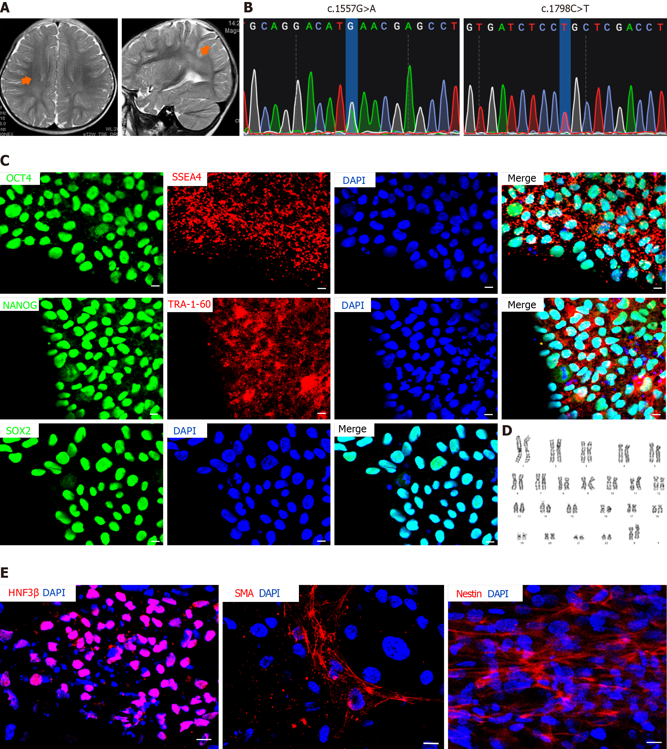
Figure 1 Generation and validation of induced pluripotent stem cell line from an infantile-onset Pompe disease patient with central nervous system involvement.
A: Cranial T2-weighted magnetic resonance imaging reveals bilateral frontoparietal white matter mild hyperintensity (orange arrows); B: Sanger sequencing confirms pathogenic variants in the acid α-glucosidase gene: C.1557G>A (p.M519I) and c.1798C>T (p.R600C); C: Immunofluorescence staining demonstrates expression of pluripotency markers (OCT4, SSEA4, NANOG, TRA1-60, and SOX2) in patient-derived induced pluripotent stem cells. Scale bar = 100 μm; D: G-banding shows a normal 46, XX karyotype in the established infantile-onset Pompe disease patient - specific induced pluripotent stem cell line; E: In vitro EB formation and immunofluorescence staining confirm trilineage differentiation potential, as evidenced by expression of hepatocyte nuclear factor 3-β (endoderm), α-smooth muscle actin (mesoderm), and Nestin (ectoderm). Scale bar = 100 μm. HNF3β: Hepatocyte nuclear factor 3-β; SMA: Smooth muscle actin.
- Citation: Jiao YC, Zhao DD, Zhang HY, Liu FC. Integrated patient induced pluripotent stem cell models and Gaa-/- mice reveal central nervous system neural pathology in infantile-onset Pompe disease. World J Stem Cells 2026; 18(7): 120578
- URL: https://www.wjgnet.com/1948-0210/full/v18/i7/120578.htm
- DOI: https://dx.doi.org/10.4252/wjsc.120578