
















Copyright: ©Author(s) 2026.
World J Stem Cells. Jul 26, 2026; 18(7): 119895
Published online Jul 26, 2026. doi: 10.4252/wjsc.119895
Published online Jul 26, 2026. doi: 10.4252/wjsc.119895
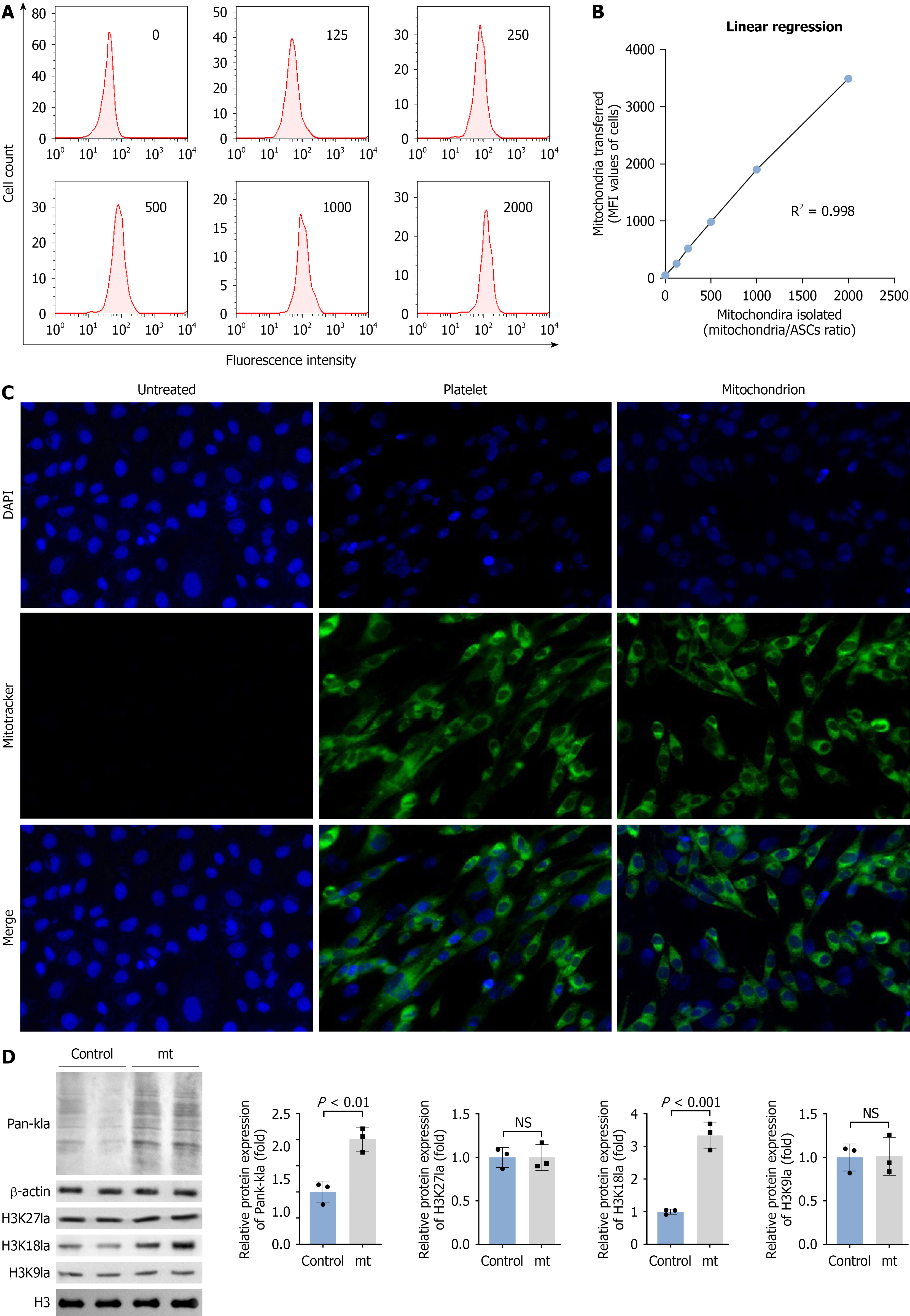
Figure 4 The transfer of mitochondria to adipose-derived stem cells promotes histone lactylation.
A: After mitochondria and adipose-derived stem cells (ASCs) were co-cultured in a series of ratios, the fluorescence intensity of mitochondrial-labeled ASCs was detected by flow cytometry; B: The linear relationship between mean fluorescence intensity value and mitochondria/ASCs ratio was analyzed using linear regression; C: In the untreated, platelet-treated and mitochondrial-treated groups of ASCs, mitochondria in ASCs were labeled with MitoTracker dye. 4’,6-diamidino-2-phenylindole was used to label the cell nuclei (200 × magnification); D: After treating with mitochondria, the levels of total lactylation (pan-kla), H3K27 lactylation (H3K27 La), H3K18 La, and H3K9 La were examined using western blotting. n = 3. Data were analyzed using Student’s t-test and are presented as mean ± SD. NS: Not significant; MFI: Mean fluorescence intensity; ASC: Adipose-derived stem cell; DAPI: 4’,6-diamidino-2-phenylindole.
- Citation: Pan Z, Zhang R, Du GM, Yi JH, Bao J, Liu CD. Platelet-derived mitochondria transfer accelerates fracture healing by promoting osteogenic differentiation and metabolic reprogramming of adipose-derived stem cells. World J Stem Cells 2026; 18(7): 119895
- URL: https://www.wjgnet.com/1948-0210/full/v18/i7/119895.htm
- DOI: https://dx.doi.org/10.4252/wjsc.119895