
















Copyright: ©Author(s) 2026.
World J Stem Cells. Jul 26, 2026; 18(7): 119260
Published online Jul 26, 2026. doi: 10.4252/wjsc.119260
Published online Jul 26, 2026. doi: 10.4252/wjsc.119260
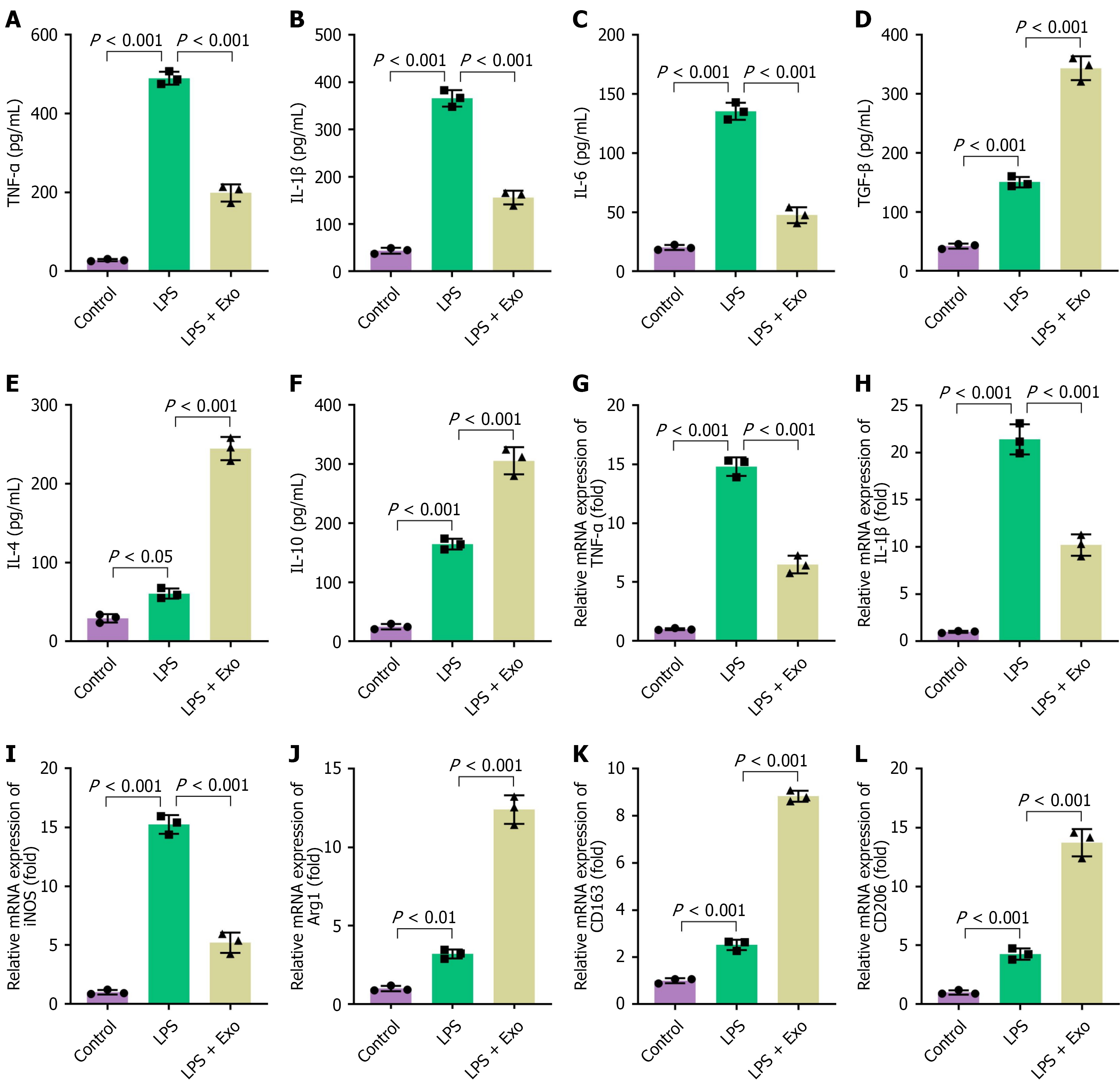
Figure 2 Exosomes modulate microglial polarization by attenuating the pro-inflammatory M1 phenotype and enhancing the anti-inflammatory M2 phenotype in vitro.
Primary microglia were stimulated with lipopolysaccharide for 24 hours and then treated with or without exosomes. A-F: The concentrations of cytokines in the culture supernatants were measured by enzyme-linked immunosorbent assay; pro-inflammatory cytokines tumor necrosis factor-alpha (A), interleukin (IL)-1β (B), and IL-6 (C); anti-inflammatory cytokines transforming growth factor-beta (D), IL-4 (E), and IL-10 (F); G-L: The mRNA expression levels of microglial polarization markers were analyzed by reverse transcription and quantitative polymerase chain reaction. M1 phenotype-related genes tumor necrosis factor-alpha (G), IL-1β (H), and inducible nitric oxide synthase (I); M2 phenotype-related genes arginase1 (J), CD163 (K), and CD206 (L). Data are presented as mean ± SD. LPS: Lipopolysaccharide; Exo: Exosome; TNF-α: Tumor necrosis factor-alpha; IL: Interleukin; TGF-β: Transforming growth factor-beta; iNOS: Inducible nitric oxide synthase; Arg1: Arginase1.
- Citation: Wang W, Wang B, Yin H. Exosomal miR-181d-5p promotes spinal cord injury repair by driving microglial polarization from M1 to M2 phenotype. World J Stem Cells 2026; 18(7): 119260
- URL: https://www.wjgnet.com/1948-0210/full/v18/i7/119260.htm
- DOI: https://dx.doi.org/10.4252/wjsc.119260