
















Copyright: ©Author(s) 2026.
World J Stem Cells. Jun 26, 2026; 18(6): 119118
Published online Jun 26, 2026. doi: 10.4252/wjsc.119118
Published online Jun 26, 2026. doi: 10.4252/wjsc.119118
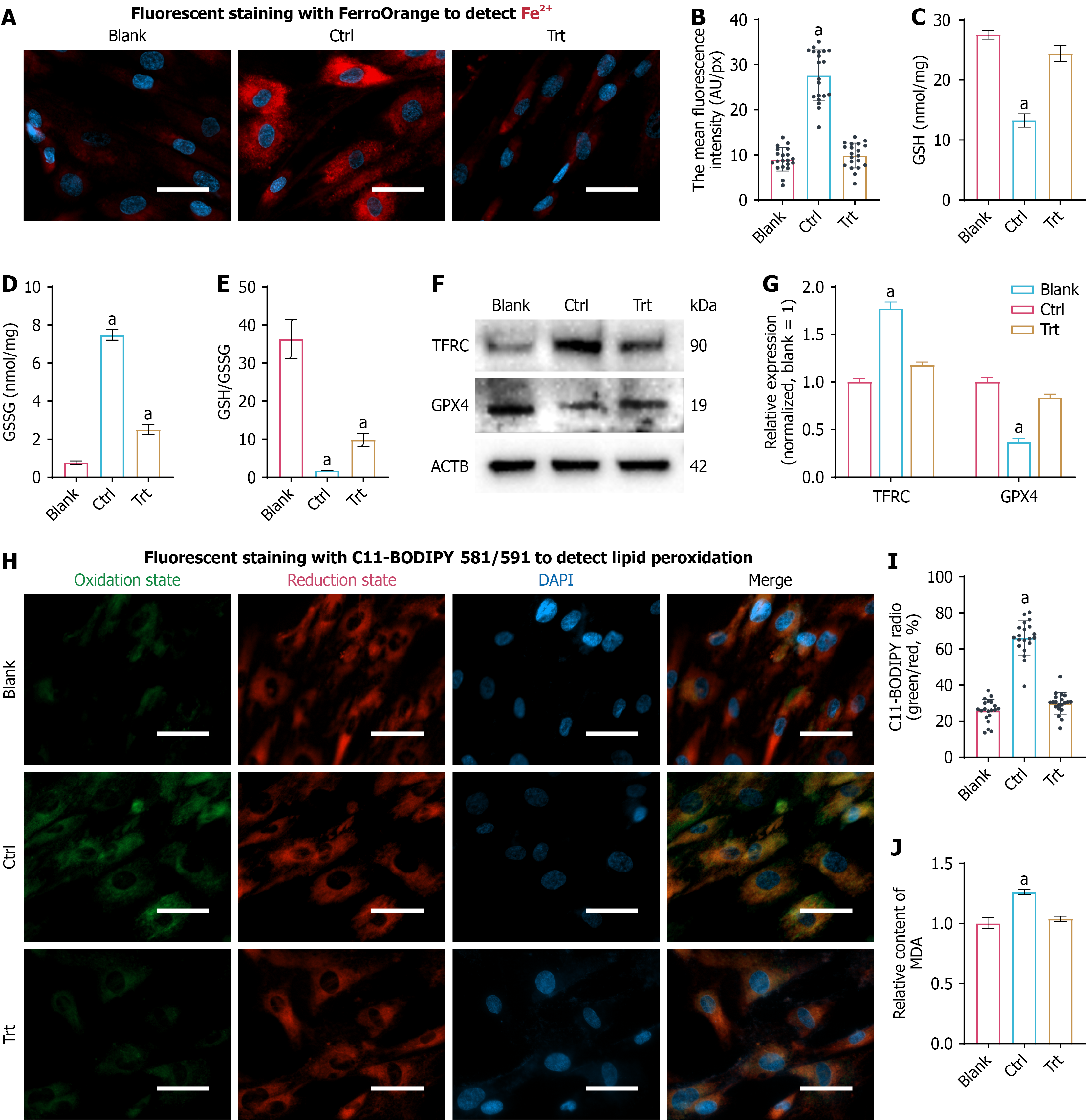
Figure 3 Selenoprotein P/selenite co-treatment restores glutathione redox balance, limits labile iron accumulation, and suppresses ferroptosis-associated lipid peroxidation.
A: Representative FerroOrange staining images showing intracellular labile Fe2+ (red) in dental pulp stem cells; nuclei were counterstained with DAPI (blue). Scale bars = 20 μm; B: Quantification of FerroOrange fluorescence intensity; C: Reduced glutathione levels; D: Oxidized glutathione levels; E: Glutathione/oxidized glutathione ratio; F: Western blot analysis of ferroptosis-associated proteins TFRC and glutathione peroxidase 4; G: Densitometric quantification of TFRC and glutathione peroxidase 4 normalized to ACTB. H: Representative C11-BODIPY 581/591 staining images for lipid peroxidation; green fluorescence indicates oxidized lipid probe and red fluorescence indicates reduced lipid probe. Scale bars = 20 μm; I: Quantification of lipid peroxidation based on the oxidized/reduced fluorescence ratio; J: Malondialdehyde levels as a biochemical readout of lipid peroxidation. Data are presented as mean ± SD. aP < 0.05. GSH: Glutathione; GSSG: Oxidized glutathione; MDA: Malondialdehyde; GPx: Glutathione peroxidase.
- Citation: Zhang RQ, Chu WH, Bai JB, Lei YH, Zhou D. Selenoprotein P attenuates oxidative senescence and ferroptosis-associated lipid peroxidation in dental pulp stem cells through a FOXM1-dependent program. World J Stem Cells 2026; 18(6): 119118
- URL: https://www.wjgnet.com/1948-0210/full/v18/i6/119118.htm
- DOI: https://dx.doi.org/10.4252/wjsc.119118