
















Copyright: ©Author(s) 2026.
World J Stem Cells. Jun 26, 2026; 18(6): 119118
Published online Jun 26, 2026. doi: 10.4252/wjsc.119118
Published online Jun 26, 2026. doi: 10.4252/wjsc.119118
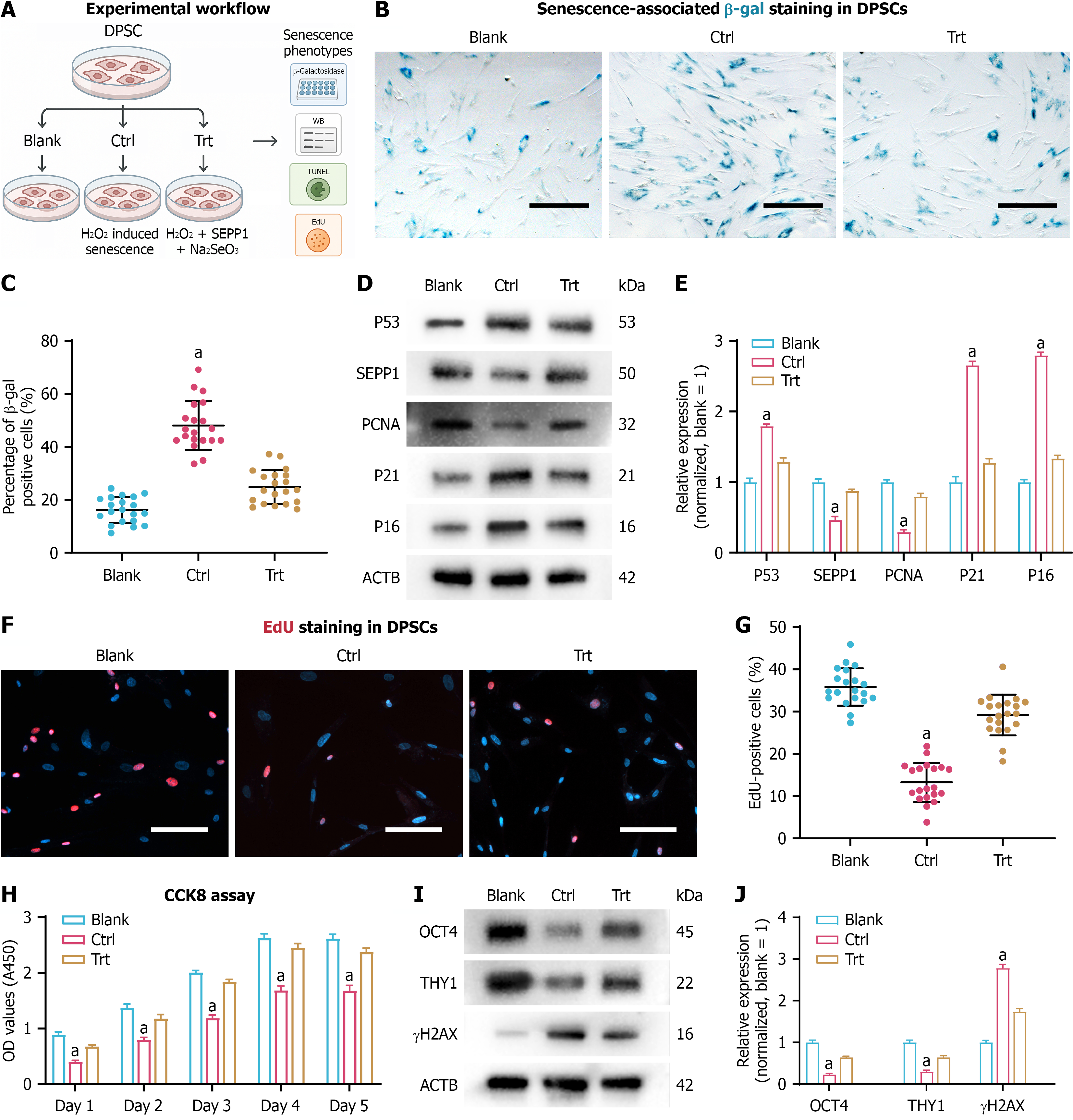
Figure 2 Selenoprotein P/selenite co-treatment attenuates H2O2-induced senescence and preserves dental pulp stem cell functional competence.
A: Experimental design for the oxidative stress-induced dental pulp stem cell (DPSC) senescence model and treatment groups [blank, control, and treatment (recombinant selenoprotein P + sodium selenite)]; B: Representative SA-β-gal staining images showing senescence-associated morphological changes in DPSCs across groups. Scale bars = 50 μm; C: Quantification of SA-β-gal-positive cells; D: Western blot analysis of senescence markers (P53, P21, and P16), selenoprotein P, and the proliferation marker proliferating cell nuclear antigen; E: Densitometric quantification of the proteins shown in panel D; F: Representative EdU staining images showing DNA synthesis/proliferation in DPSCs. Scale bars = 50 μm; G: Quantification of the percentage of EdU-positive cells; H: CCK-8 assay showing growth curves/viability of DPSCs across groups; I: Western blot analysis of stemness markers (OCT4 and THY1) and the DNA damage marker γH2AX; J: Densitometric quantification of OCT4, THY1, and γH2AX. Data are presented as mean ± SD. aP < 0.05. DPSC: Dental pulp stem cell; SEPP1: Selenoprotein P; PCNA: Proliferating cell nuclear antigen.
- Citation: Zhang RQ, Chu WH, Bai JB, Lei YH, Zhou D. Selenoprotein P attenuates oxidative senescence and ferroptosis-associated lipid peroxidation in dental pulp stem cells through a FOXM1-dependent program. World J Stem Cells 2026; 18(6): 119118
- URL: https://www.wjgnet.com/1948-0210/full/v18/i6/119118.htm
- DOI: https://dx.doi.org/10.4252/wjsc.119118