
















Copyright: ©Author(s) 2026.
World J Stem Cells. Jun 26, 2026; 18(6): 117622
Published online Jun 26, 2026. doi: 10.4252/wjsc.117622
Published online Jun 26, 2026. doi: 10.4252/wjsc.117622
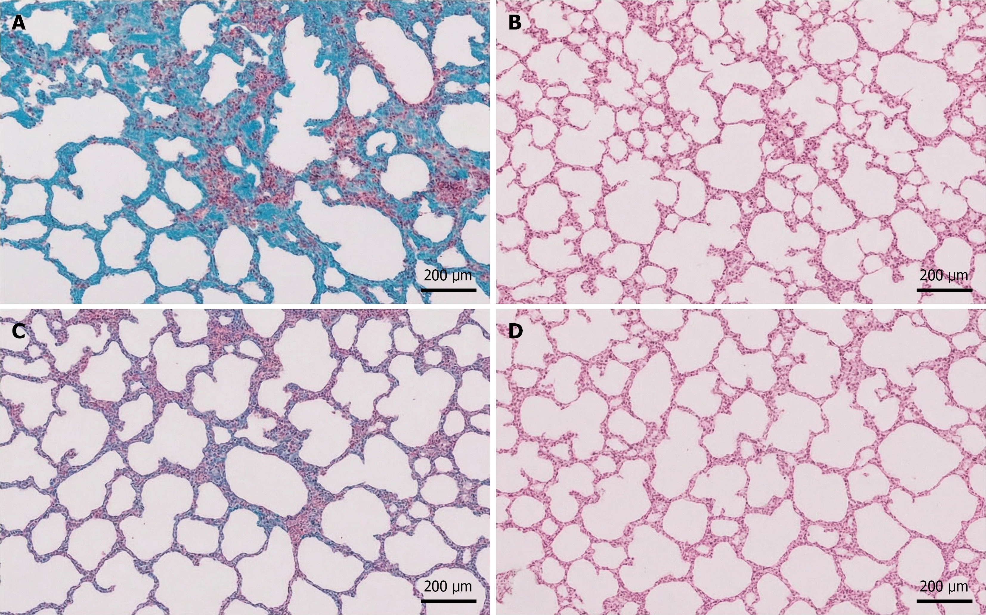
Figure 3 Histopathological assessment of pulmonary fibrosis and inflammation.
A-D: Lung tissue sections stained with Masson’s trichrome and hematoxylin and eosin were evaluated on the 28th day. In the model group, extensive blue-stained collagen deposition, disrupted alveolar architecture, and prominent inflammatory cell infiltration were observed (A). In the mitoquinone group (B) and mesenchymal stem cells group (C), collagen deposition and inflammatory infiltration were reduced. In the combination group, lung tissue architecture was largely preserved, with minimal collagen deposition and inflammatory cell infiltration, indicating a significant synergistic effect of the combination treatment in attenuating pulmonary fibrosis and inflammation (D), scale bar = 200 μm.
- Citation: Hu YL, Lin ZX, Zhang XY, Li GL, Liu WJ, Shi ZQ, Li XH, Yang ZZ. Combining mitochondrial antioxidant mitoquinone and mesenchymal stem cell therapy for amelioration of radiation-induced lung injury: Synergistic regenerative strategy. World J Stem Cells 2026; 18(6): 117622
- URL: https://www.wjgnet.com/1948-0210/full/v18/i6/117622.htm
- DOI: https://dx.doi.org/10.4252/wjsc.117622