
















Copyright: ©Author(s) 2026.
World J Stem Cells. Jun 26, 2026; 18(6): 117622
Published online Jun 26, 2026. doi: 10.4252/wjsc.117622
Published online Jun 26, 2026. doi: 10.4252/wjsc.117622
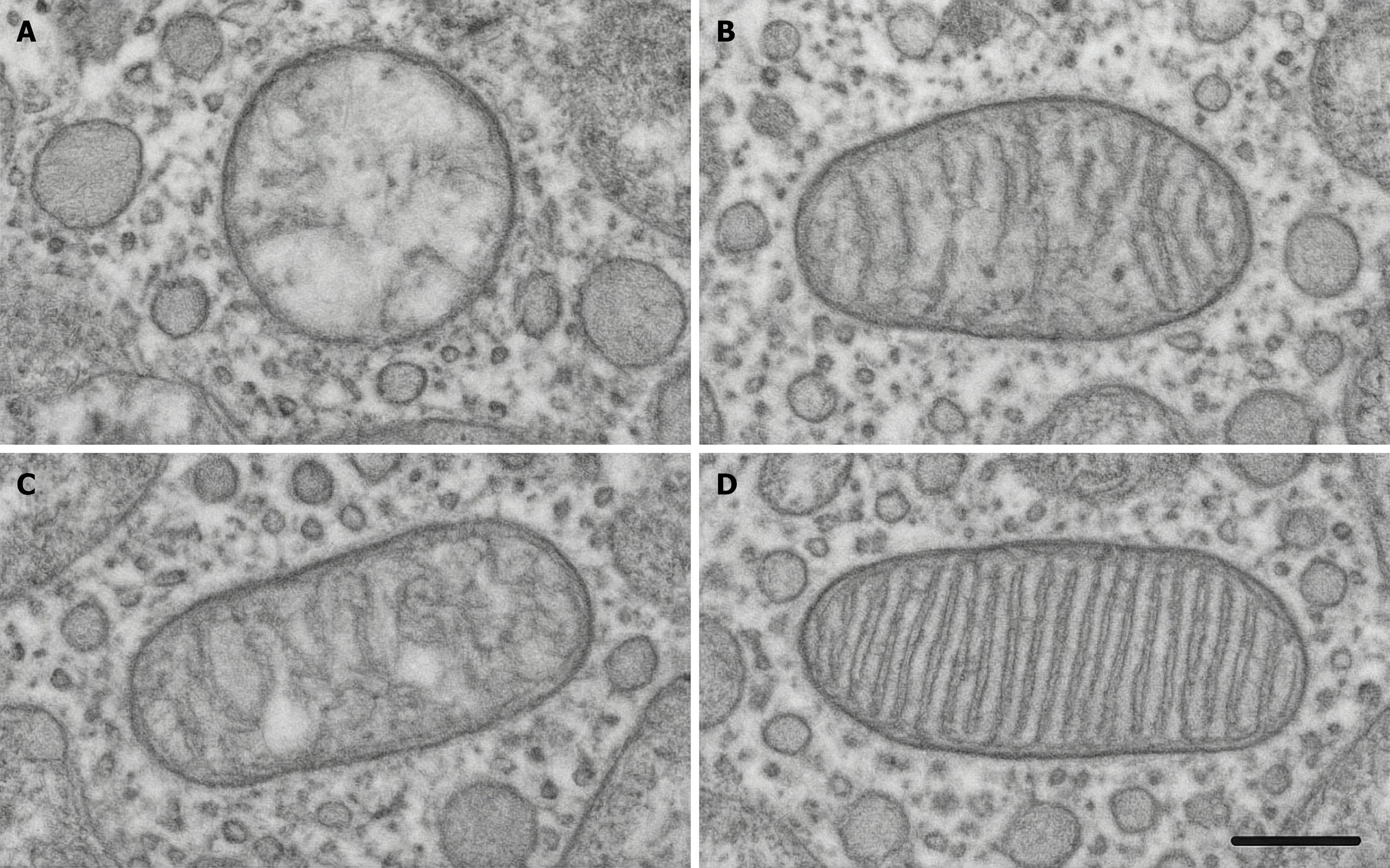
Figure 2 Transmission electron microscopy of mitochondrial ultrastructure.
A-D: Mitochondrial ultrastructure in lung tissue was examined by transmission electron microscopy. In the model group, mitochondria exhibited pronounced swelling, cristae disruption or loss, and indistinct membrane structures (A). In the mitoquinone group (B) and the mesenchymal stem cells group (C), mitochondrial morphology was partially restored, with relatively preserved cristae. The combination group displayed the most intact mitochondrial morphology, with well-organized cristae closely resembling normal structure, suggesting superior protection of mitochondrial structural integrity by the combination treatment (D), scale bar = 0.5 μm.
- Citation: Hu YL, Lin ZX, Zhang XY, Li GL, Liu WJ, Shi ZQ, Li XH, Yang ZZ. Combining mitochondrial antioxidant mitoquinone and mesenchymal stem cell therapy for amelioration of radiation-induced lung injury: Synergistic regenerative strategy. World J Stem Cells 2026; 18(6): 117622
- URL: https://www.wjgnet.com/1948-0210/full/v18/i6/117622.htm
- DOI: https://dx.doi.org/10.4252/wjsc.117622