
















Copyright: ©Author(s) 2026.
World J Stem Cells. Jun 26, 2026; 18(6): 117622
Published online Jun 26, 2026. doi: 10.4252/wjsc.117622
Published online Jun 26, 2026. doi: 10.4252/wjsc.117622
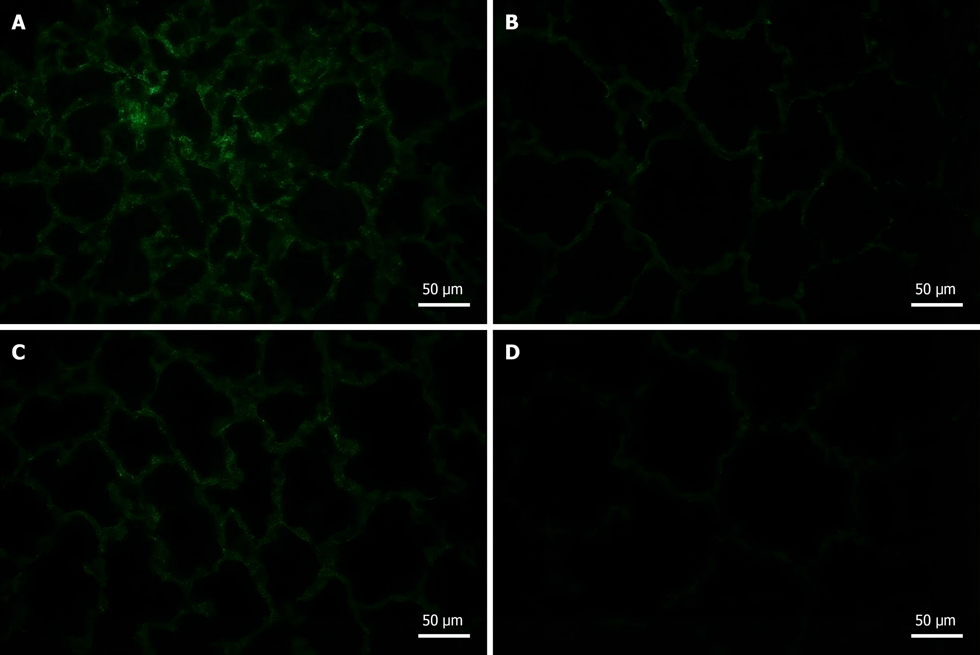
Figure 1 Oxidative stress levels in lung tissue.
A-D: Representative fluorescence microscopic images of reactive oxygen species (ROS) staining in lung tissue of each group were obtained on day 28. In the model group (A), diffuse and intense fluorescence was observed, indicating marked ROS accumulation. In the mitoquinone group (B) and the mesenchymal stem cells group (C), fluorescence intensity was significantly reduced and more localized. The combination group exhibited the weakest fluorescence signal, approaching that of normal lung tissue, indicating a synergistic effect of the combination treatment in reducing ROS levels (D), scale bar = 50 μm.
- Citation: Hu YL, Lin ZX, Zhang XY, Li GL, Liu WJ, Shi ZQ, Li XH, Yang ZZ. Combining mitochondrial antioxidant mitoquinone and mesenchymal stem cell therapy for amelioration of radiation-induced lung injury: Synergistic regenerative strategy. World J Stem Cells 2026; 18(6): 117622
- URL: https://www.wjgnet.com/1948-0210/full/v18/i6/117622.htm
- DOI: https://dx.doi.org/10.4252/wjsc.117622