
















Copyright: ©Author(s) 2026.
World J Stem Cells. Jun 26, 2026; 18(6): 116526
Published online Jun 26, 2026. doi: 10.4252/wjsc.116526
Published online Jun 26, 2026. doi: 10.4252/wjsc.116526
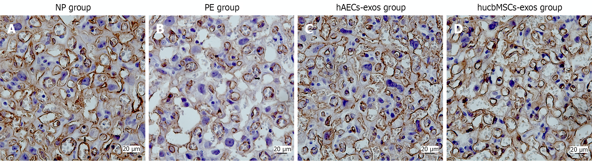
Figure 13 Immunohistochemical staining of placental tissues from rats (400 ×).
A: Immunohistochemical staining results of CD31-positive cells in the placenta of rats in the normal pregnancy group; B: Immunohistochemical staining results of CD31-positive cells in the placenta of rats in the preeclampsia group; C: Immunohistochemical staining results of CD31-positive cells in the placenta of rats in the human amniotic epithelial cell exosomes group; D: Immunohistochemical staining results of CD31-positive cells in the placenta of rats in the exosomes from mesenchymal stem cells derived from human umbilical cord blood group. NP: Normal pregnancy; PE: Preeclampsia; hAECs-exos: Human amniotic epithelial cell exosomes; hucbMSCs-exos: Exosomes from mesenchymal stem cells derived from human umbilical cord blood.
- Citation: Han LY, Zhang WZ, Zhang Y, Zhong W, Sun JL, Shen J. Therapeutic effects of human amniotic epithelial cell-derived and umbilical cord blood mesenchymal stem cell-derived exosomes on preeclamptic rats. World J Stem Cells 2026; 18(6): 116526
- URL: https://www.wjgnet.com/1948-0210/full/v18/i6/116526.htm
- DOI: https://dx.doi.org/10.4252/wjsc.116526