











BPG is committed to discovery and dissemination of knowledge
Opinion Review
Copyright: ©Author(s) 2026.
World J Stem Cells. May 26, 2026; 18(5): 119231
Published online May 26, 2026. doi: 10.4252/wjsc.v18.i5.119231
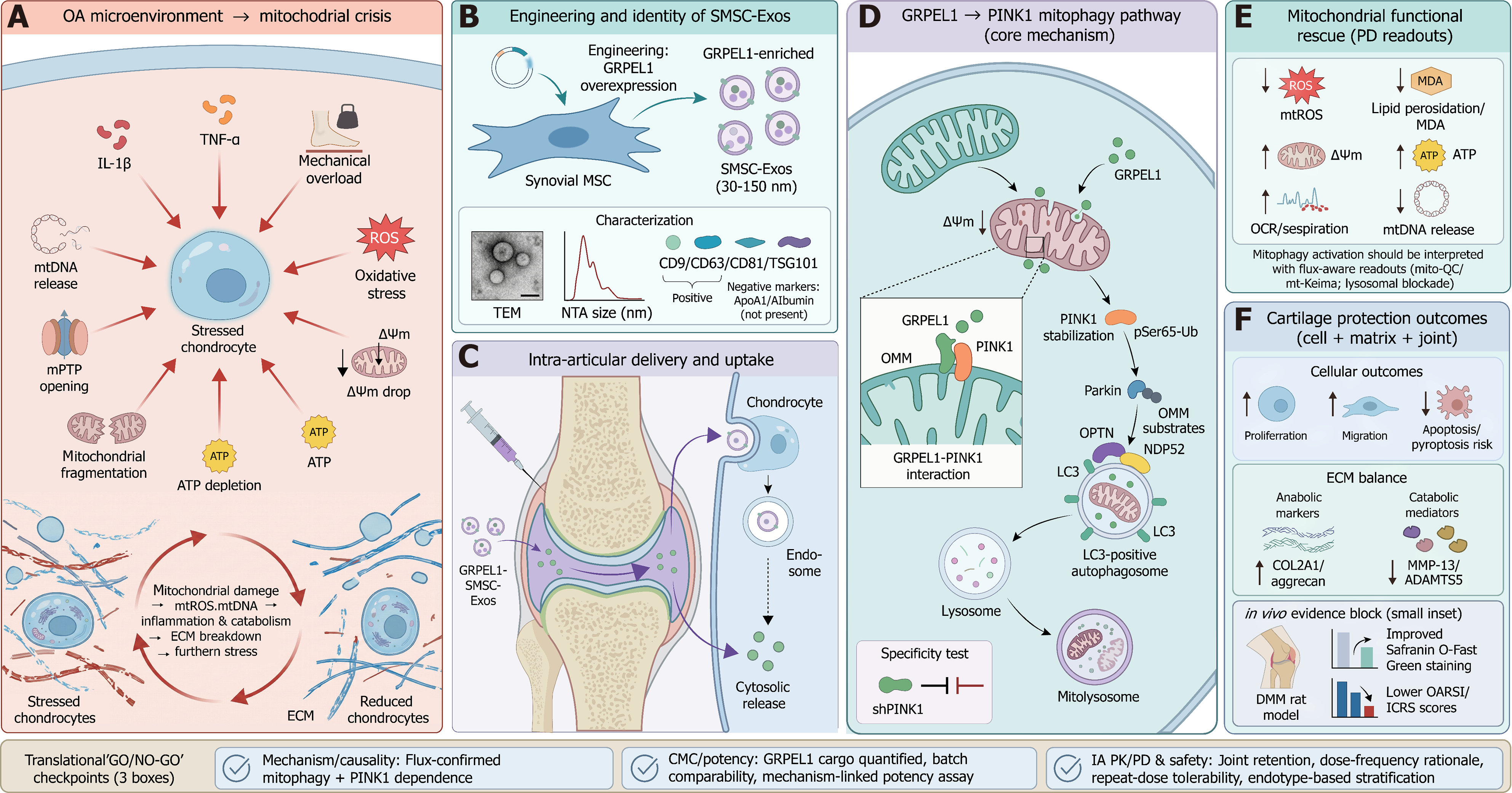
Figure 1
Figure 1 GrpE-like 1-loaded synovial mesenchymal stem cell-derived exosomes activate phosphatase and tensin homolog-induced kinase 1-dependent mitophagy to protect cartilage in osteoarthritis. A: Osteoarthritis microenvironment-mitochondrial crisis. Inflammatory cytokines (interleukin-1β and tumor necrosis factor-alpha) and mechanical overload converge on chondrocytes to trigger oxidative stress [reactive oxygen species (ROS)], mitochondrial fragmentation, mitochondrial permeability transition pore opening, mitochondrial DNA (mtDNA) release, mitochondrial membrane potential (ΔΨm) loss, and ATP depletion, forming a self-amplifying loop of mitochondrial damage → mitochondrial ROS/mtDNA → inflammation/catabolism → extracellular matrix breakdown → further stress; B: Engineering and identity of synovial mesenchymal stem cell-derived exosomes. Synovial mesenchymal stem cells are engineered to overexpress GrpE-like 1 (GRPEL1), generating GRPEL1-enriched exosomes (30-150 nm). Exosomes are characterized by morphology (transmission electron microscopy), size distribution (nanoparticle tracking analysis), extracellular vesicle markers (CD9/CD63/CD81/tumor susceptibility gene 101), and absence of selected impurities/negative markers (apolipoprotein A1/albumin); C: Intra-articular delivery and uptake. GRPEL1-synovial mesenchymal stem cell-derived exosomes are administered by intra-articular injection, traffic within the joint space, and are internalized by chondrocytes via endocytosis followed by intracellular release of functional cargo; D: Core mechanism: GRPEL1 → phosphatase and tensin homolog-induced kinase 1 (PINK1) mitophagy pathway. GRPEL1 localizes to mitochondria and associates with PINK1 at the outer mitochondrial membrane, promoting PINK1 stabilization, increased Ser65-phosphorylated ubiquitin (pSer65-Ub), Parkin recruitment, ubiquitination of outer mitochondrial membrane substrates, adaptor recruitment (optineurin/NDP52), LC3-positive autophagosome formation, and lysosomal fusion to generate mitolysosomes for mitochondrial clearance. A pathway specificity inset illustrates inhibition of downstream events by PINK1 knockdown (shPINK1); E: Mitochondrial functional rescue (pharmacodynamics readouts). Pathway engagement is associated with reduced mitochondrial ROS and lipid peroxidation (malondialdehyde), improved ΔΨm, increased ATP and respiratory activity (oxygen consumption rate), and reduced mtDNA release. Mitophagy activation should be interpreted with flux-aware readouts (e.g., mito-QC/mt-Keima reporters and/or lysosomal end-blockade designs); F: Cartilage protection outcomes across scales. At the cellular level, GRPEL1-exosomes support proliferation and migration while reducing apoptosis/pyroptosis risk. At the matrix level, anabolic markers (collagen type II alpha 1/aggrecan) increase and catabolic mediators (matrix metalloproteinase-13/A disintegrin and metalloproteinase with thrombospondin motifs 5) decrease. In vivo (destabilization of the medial meniscus rat osteoarthritis model), cartilage protection is reflected by improved Safranin O-Fast Green staining and lower Osteoarthritis Research Society International/International Cartilage Repair Society scores. The footer summarizes translational “Go/No-Go” checkpoints spanning: (1) Mechanism/causality (flux-confirmed, PINK1-dependent mitophagy); (2) Chemistry, manufacturing, and controls/potency (cargo quantification, batch comparability, mechanism-linked potency assay); and (3) Intra-articular pharmacokinetics/pharmacodynamics and safety (joint retention, dose-frequency rationale, repeat-dose tolerability, and endotype-based stratification). OA: Osteoarthritis; IL-1β: Interleukin-1β; TNF-α: Tumor necrosis factor-alpha; mtDNA: Mitochondrial DNA; mPTP: Mitochondrial permeability transition pore; ΔΨm: Mitochondrial membrane potential; mtROS: Mitochondrial reactive oxygen species; ECM: Extracellular matrix; SMSC-Exos: Synovial mesenchymal stem cell-derived exosomes; GRPEL1: GrpE-like 1; MSC: Mesenchymal stem cell; TEM: Transmission electron microscopy; NTA: Nanoparticle tracking analysis; ApoA1: Apolipoprotein A1; PINK1: Phosphatase and tensin homolog-induced kinase 1; OMM: Outer mitochondrial membrane; OPTN: Optineurin; PD: Pharmacodynamics; ROS: Reactive oxygen species; MDA: Malondialdehyde; OCR: Oxygen consumption rate; COL2A1: Collagen type II alpha 1; MMP-13: Matrix metalloproteinase-13; ADAMTS5: A disintegrin and metalloproteinase with thrombospondin motifs 5; OARSI: Osteoarthritis Research Society International; ICRS: International Cartilage Repair Society; CMC: Chemistry, manufacturing, and controls; MQC: Mitochondrial quality control; IA: Intra-articular; PK: Pharmacokinetics.


Write to the Help Desk