
















Copyright: ©Author(s) 2026.
World J Stem Cells. May 26, 2026; 18(5): 118307
Published online May 26, 2026. doi: 10.4252/wjsc.v18.i5.118307
Published online May 26, 2026. doi: 10.4252/wjsc.v18.i5.118307
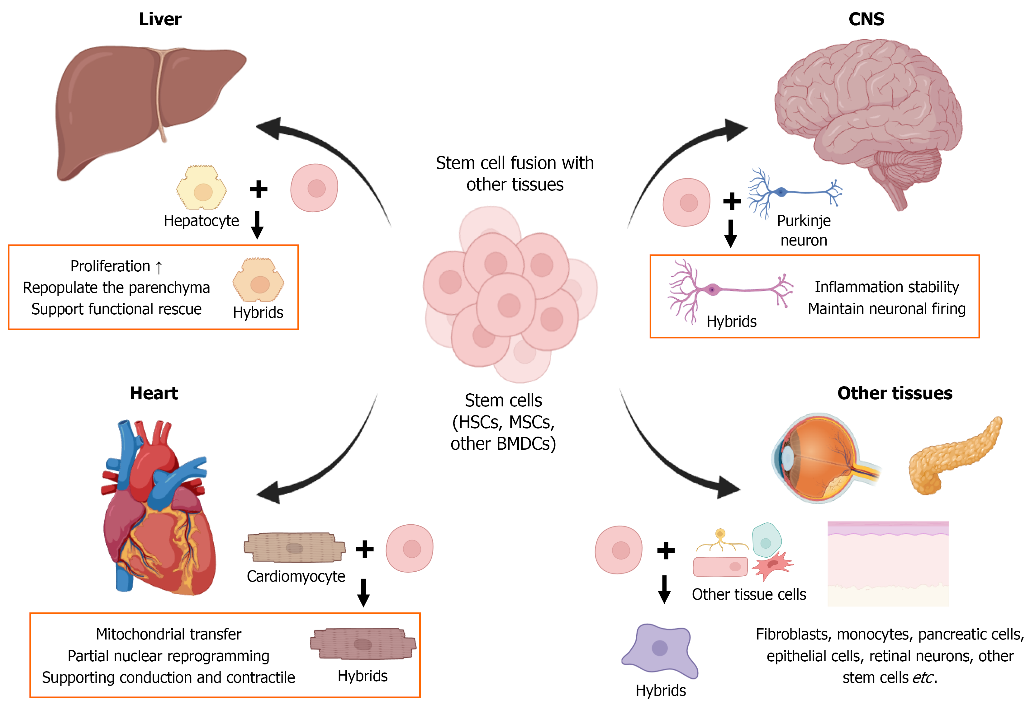
Figure 3 Stem cell fusion-mediated regeneration in diverse tissues.
Stem cells and bone marrow-derived cells (BMDCs) can contribute to tissue repair not only through paracrine signaling and differentiation but also by fusing with host somatic cells in injured organs. In this schematic, a central node representing hematopoietic stem cells, mesenchymal stromal/stem cells, and other BMDCs is connected to multiple target tissues. In the liver, BMDC-hepatocyte fusion generates hybrid cells that can proliferate, repopulate the parenchyma, and support functional rescue in transplantation models. In the central nervous system, BMDCs fuse with Purkinje neurons to form binucleated heterokaryons that maintain neuronal firing properties and may contribute to circuit stability in the context of inflammation. In the heart, fusion between stem cells and cardiomyocytes can facilitate mitochondrial transfer, partial nuclear reprogramming, and integration into contractile syncytia, thereby supporting conduction and contractile function. In skeletal muscle, satellite cells and, under some conditions, BMDCs fuse with damaged myofibers to restore multinucleated fibers and maintain muscle architecture. Fusion events have also been reported in other tissues, including epithelium, retina, and pancreas, where their contribution to long-term regeneration remains to be fully defined. Across these contexts, tissue injury, inflammation, chemokine gradients, and extracellular matrix remodeling create fusion-permissive niches that recruit and prime stem cells to engage in fusion-mediated repair. CNS: Central nervous system; HSCs: Hematopoietic stem cells; MSCs: Mesenchymal stromal/stem cells; BMDCs: Bone marrow-derived cells.
- Citation: Kim JS, Hwang IS, Kim SJ, Moon MH, Park HD, Jin BH, Kim DY, Lee MJ, Eum SY, Yoo SJ, Cha BH. Cell-cell fusion and stem cell plasticity: Emerging paradigms in tissue regeneration. World J Stem Cells 2026; 18(5): 118307
- URL: https://www.wjgnet.com/1948-0210/full/v18/i5/118307.htm
- DOI: https://dx.doi.org/10.4252/wjsc.v18.i5.118307